Our group studies the spatial architectures and phenotypes of cells and tissues under different human diseases and environmental influences. We develop and use quantitative imaging assays and machine learning models to predict the biological effects of genetic mutations, drugs, and/or environmental agents. Our current research areas include spatial profiling of cells and tissues, bioimage databases and portals, digital medicine for cancer, and toxicodynamics of drugs/chemicals (Fig. 1).
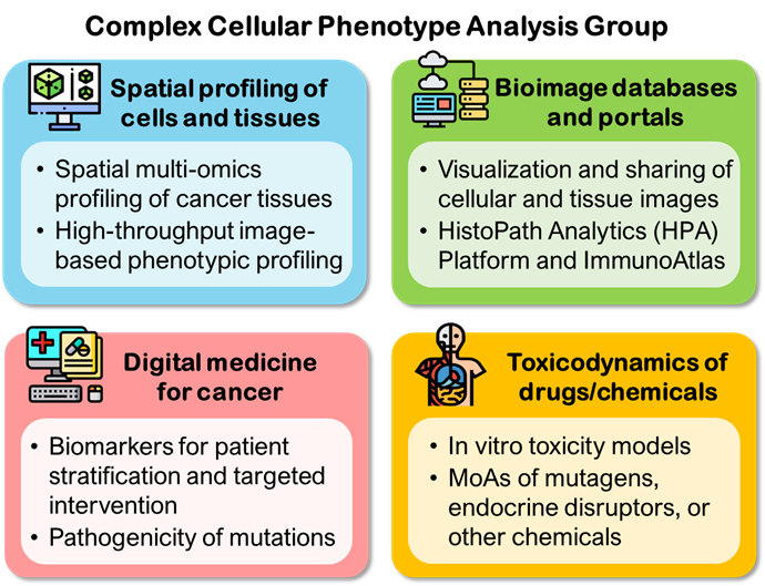
Figure 1. Our current research areas
Our members come from diverse scientific backgrounds, including chemistry, cell biology, immunology, computer science, and bioinformatics. We collaborate with different academic, clinical, industrial, and governmental research groups, including Institute of Molecular and Cell Biology (IMCB), Singapore Institute of Food and Biotechnology Innovation (SIFBI), Singapore General Hospital (SGH), National Cancer Centre Singapore (NCCS), Lee Kong Chain (LKC) School of Medicine, and Harvard Beth Israel Deaconess Medical Center (BIDMC).
Recent advances in multiplex immunohistochemistry/immunofluorescence (mIHC/IF) technologies have enabled simultaneous measurements of large numbers of markers on the same tissue sections, and more comprehensive views of the cellular compositions and immune responses at the tumor microenvironment (TME). We have developed computational methods and software tools to construct quantitative and compact representations of cellular or tissue phenotypes based on these multiplexed cellular or tissue images (Fig. 2). Our methods and tools can handle terabytes of image data collected under large numbers of experimental conditions or patients. Phenotypic profiles constructed using these methods have been used to classify the effects of small molecules and assess potentially harmful effects of chemicals and environmental agents (Hussain et al., 2020; Friedman et al., 2019; Lee et al., 2018; and Su et al., 2016).
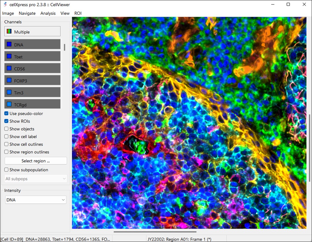
Figure 2. CellXpress 2.0 can handle and quantify highly-multiplexed and large human tissue images.
The reproducibility and interpretation of the complex staining patterns and analysis results obtained from mIHC/IF technologies are vital to their general adoptions. We develop and maintain an online platform for managing, visualizing, and sharing large tissue images called the HistoPathology Analytics (HPA) Platform (Fig. 3). The platform can help researchers and clinicians to more rapidly and accurately quantify the effects of cancer therapeutic agents (Yeong et al., 2022, Leong et al., 2021). An online public portal for mIHC/IF images and results for immuno-oncology called ImmunoAtlas (https://ImmunoAtlas.org) has also been built based on the HPA Platform (Lee et al., 2021).
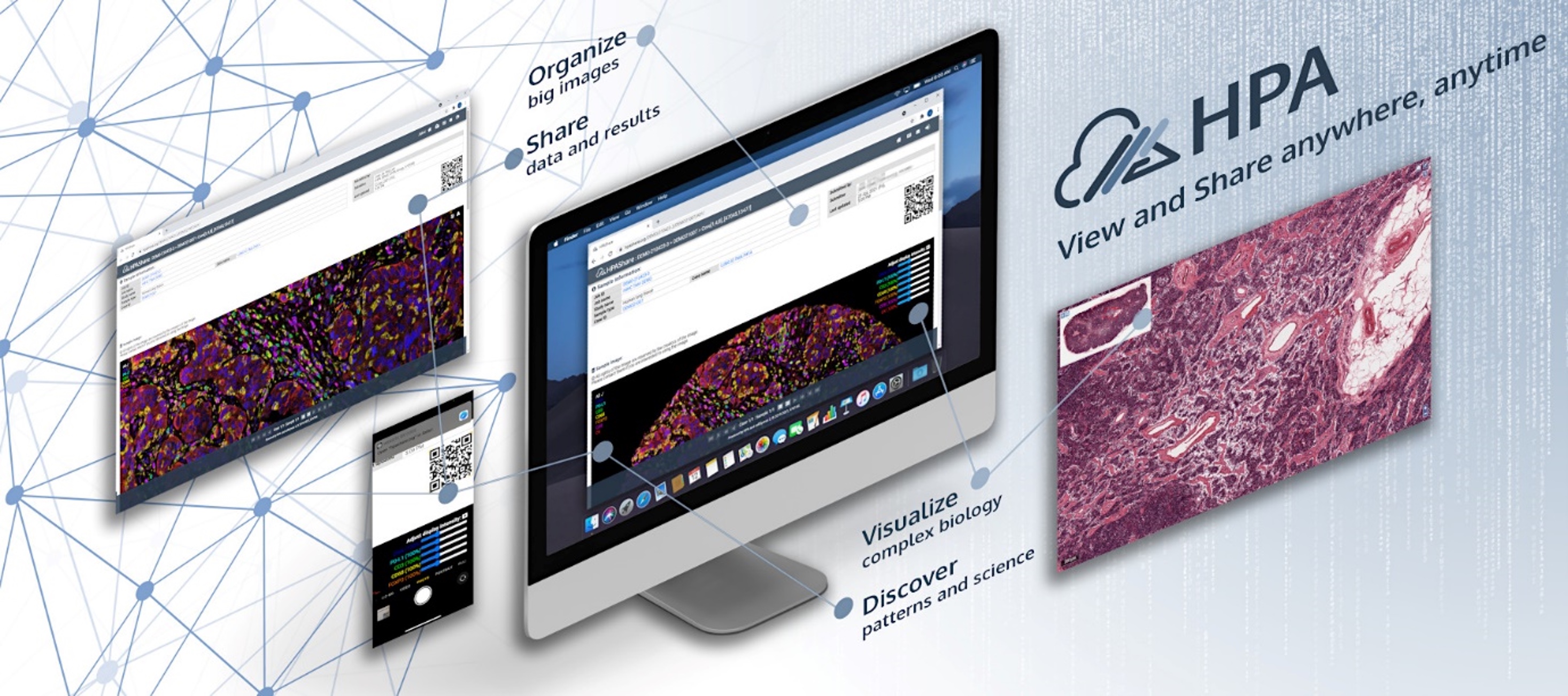
Figure 3. HistoPath Analytics (HPA) is a cloud-based digital histopathology platform for organizing, sharing, visualizing, and analyzing large histological images.
Recent advances in genomic, transcriptomic, phenotypic, and histopathological profiling technologies have enabled the generation histopathological profiling technologies have enabled the generation of large amounts of molecular and phenotypic information about the physiopathology of individual cancer patients. We are developing machine learning and data analytics methods to integrate data from these diverse technologies to stratify patients and select optimum targeted interventions for hepatocellular carcinoma, breast and other cancers. We are also developing imaging assays that can predict the pathogenicity of genetic variants of key cancer-associated genes.
Many xenobiotics have unknown and/or non-specific intracellular targets. To study the toxicodynamics of these chemicals, unbiased approaches that do not require prior information about the targets or mechanisms of the chemicals are required. Our focus is to study chemical analogs with related structures but differential cellular effects (Goh et al., 2021; Jaladanki et al., 2021), and develop fit-for-purpose assays that will be used by regulatory agencies and industrial research laboratories to assess chemical safety. We have developed a high-throughput and predictive in vitro pulmonary toxicity assay based on a human bronchial epithelial cell line, BEAS-2B (Fig. 4; Lee et al., 2018). The assay can accurately classify 33 reference chemicals with human pulmonotoxicity information (88.8% balance accuracy, 84.6% sensitivity, and 93.0% specificity). We also participated in an international case study that demonstrates the utility of in vitro bioactivity as a lower bound estimate of in vivo adverse effect levels in risk-based prioritization (Friedman et al., 2020).
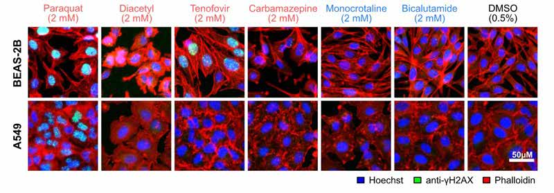
Figure 4. Immunofluorescence microscopy images of human lung cells showing different phenotypic responses to non-toxic (blue) and toxic (red) chemicals.
| Senior Principal Investigator | LOO Lit Hsin | [View Bio] |
| Lead Research Officer | LEE Jia Ying Joey |
| Senior Research Officer | KONG Jia Wen Carmen |
| Research Officer | GONZALES Edward Mark |
| PhD Student | YEO Chyi Maey Claresta |
 | LOO Lit Hsin Senior Principal Investigator Email: loolh@a-star.edu.sg Research Group: Complex Cellular Phenotype Analysis |
Dr. Loo Lit Hsin is a Senior Principal Investigator at the Bioinformatics Institute (BII), A*STAR, Singapore. He is also an adjunct Assistant Professor at the Department of Pharmacology, Yong Loo Lin School of Medicine, National University of Singapore. Dr. Loo’s background is in computational and systems pharmacology/toxicology. He was the recipient of the Lush Prize – Science Award (2016), Award for Excellence in Postdoctoral Research (2010) and the Alfred Gilman Award (2009) by the University of Texas Southwestern (UTSW) Medical Center, and the Doctoral Award in Mathematical Sciences and Engineering (2005) by Drexel University. Dr. Loo was a postdoctoral fellow in the Bauer Center for Genomics Research at Harvard University (2005), and then in the Department of Pharmacology at the UTSW Medical Center, USA (2005-2010).
| Lead Research Officer | LEE Jia Ying Joey |
| Senior Research Officer | KONG Jia Wen Carmen |
| Research Officer | GONZALES Edward Mark |
| PhD Student | YEO Chyi Maey Claresta |