A*STAR NEWS
PRESS RELEASES
Singapore Scientists Discover Human Liver Cancer Cells Masquerading as Fetal-Like Cells
Liver cancers adopt a fetal-like environment to escape immune surveillance and grow more aggressively

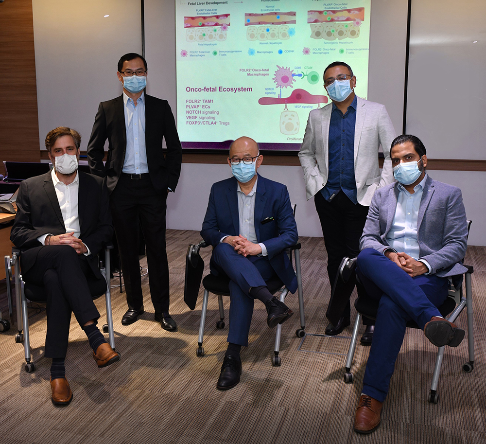
From left: Dr Florent Ginhoux, Prof Jerry Chan, Prof Pierce Chow, Dr Ramanuj DasGupta, and Dr Ankur Sharma (Copyright: A*STAR’s Genome Institute of Singapore)
SINGAPORE – A team of researchers and clinician-scientists from A*STAR’s Genome Institute of Singapore (GIS), Singapore Immunology Network (SIgN), National Cancer Centre Singapore (NCCS), and KK Women’s and Children’s Hospital (KKH), have identified a pivotal “fetal-like” reprogramming of the tumour ecosystem in human hepatocellular carcinoma (HCC). This study leveraged the platform established by the National Medical Research Council (NMRC) Translational and Clinical Research (TCR) Flagship Programme in Liver Cancer1 and was recently published in Cell on 24 September 2020.
HCC, or primary liver cancer, is the sixth most common cancer in the world, and the second most common cause of cancer deaths globally2. It is estimated that approximately 80% of all HCC are found in Asia. Tumours represent a complex ecosystem of diverse cell types that exhibit molecular and functional heterogeneity and plasticity. Previous studies have noted the similarities between embryonic development and tumours, specifically in the expression of fetal-like antigens in cancers. Most notably, alpha-fetoprotein (AFP) is one of the key onco-fetal proteins found to be elevated in more than 80% of HCC, especially in those with poorly differentiated cell types which are clinically more aggressive. The fetal immune system and placental-fetal system are known to be largely immunologically tolerant, allowing their co-existence within the mother until birth. It has long been suggested that cancers may have similar mechanisms to avoid rejection by the body. While it is known that HCC can undergo onco-fetal reprogramming, the relationship between HCC and its micro-environment has not been examined in detail.
In this comparative study, the researchers mapped approximately 200,000 single cells in the human liver across the timeline from fetal development to terminal liver cancer. It led to the discovery of two new fetal-associated cell types in the tumour ecosystem in HCC. Specifically, the team identified the reappearance of fetal-like endothelial cells that are responsible for blood supply, and fetal-like macrophages, an immune cell type that serves as the body’s first line of defence against pathogens, but are also associated with tissue homeostasis.
The researchers discovered that during development of the fetal liver (a process that shares many characteristics typically associated with cancer growth), these cell types (fetal endothelial and macrophages) help in organ growth and protect the tissue against the body’s own immune system. The team identified the sequential activation of VEGF and Notch signalling pathways that led to fetal-like reprogramming of endothelial cells and macrophages. Importantly, the authors showed that these signalling pathways are activated during fetal-liver growth, and switched off thereafter, but are re-activated during the development of liver cancer.
A dividing cancer cell first activates VEGF pathway in endothelial cells, which in turn communicate with macrophages in tumours via Notch-Delta interactions. The fetal-like programme in tumours allows uncontrolled growth and protection from the immune system. This study provides unprecedented and fundamental insights into the processes that drive cancer development, and may provide new therapeutic strategies to re-activate the immune system in the fight against cancer.
Dr Ramanuj DasGupta, cancer biologist and Senior Group Leader at GIS, and the lead corresponding author of the study, said, “Our research underscores a fundamental understanding of how early developmental processes can be hijacked by cancers in order to facilitate their own growth, and escape immune surveillance. This study also opens up exciting possibilities for the discovery of biomarkers and novel therapeutic targets associated with non-malignant cells, especially in the context of combinatorial immunotherapy with anti-angiogenic drugs. Most importantly, it highlights the power of likeminded scientists and clinicians coming together to solve big problems. I am absolutely thrilled to be working with this dream-team of exceptional scientists and clinicians.”
Dr Ankur Sharma, Research Scientist at GIS, the first and co-corresponding author of the study, said, “Our research provides insights into the mirror worlds of fetal and tumour ecosystem, where certain cell types first appear during organ development and later reappear in cancer. The ‘reappearance’ of fetal-like cells provides the perfect environment for tumour growth by supressing the immune system. Our work demonstrates the importance of embryonic macrophages in tumour growth and immune suppression. This study opens new avenues to understand the embryonic origin of cancers and its implication in discovering new therapeutic approaches, especially in the realm of immunotherapy.”
Dr Florent Ginhoux, scientist and Senior Principal Investigator at SIgN, and cocorresponding author of the study said, “The macrophages are one of the first immune cells to arise during development as their raison d’être is to provide adequate organ building and tissue maintenance. These embryonic macrophages are endowed with immunesuppressive properties as inflammation too early would be detrimental to the correct development of fetal tissues. Our work uncovers that tumour cells exploit embryonic macrophage’s raison d’être for their own growth, by recreating an artificial fetal-like environment and taking macrophages ‘back in time’ mimicking early developmental settings. This study will help to understand the importance of macrophage ontogeny in cancer development and open new strategies to eradicate cancer.”
Prof Pierce Chow, Clinician-Scientist and Liver Surgeon at NCCS, Principal Investigator of the NMRC TCR Flagship Programme in Liver Cancer, and co-corresponding author of the study, said, “This study provides mechanistic confirmation of what many liver cancer specialists have suspected – regression to a fetal state allows HCC to grow aggressively. The findings potentially opens up new approaches in the prevention and treatment of HCC. It is also a showcase of what multi-disciplinary collaborative research between clinicianscientists from different specialities and scientists with different expertise can achieve.”
Prof Chow who is also a Programme Director with Duke-NUS Medical School added that he may include the investigation for proteins uncovered by this study in a prospective cohort study of patients at high risk of developing liver cancer, which has been planned to start at the end of 2020.
Prof Jerry Chan, Clinician-Scientist and Senior Consultant at KKH’s Department of Reproductive Medicine and co-author of the study, said, “Globally, cancer cases have risen rapidly over the years. Understanding the behaviour of how cancer cells reprogramme their environment to simulate a fetal environment, and thereby promoting their growth and avoiding immune destruction, may be key to developing a cure. KKH is proud to contribute to this novel discovery through underlying expertise in the developing fetal immune system3, in collaboration with A*STAR and others."
Prof William Hwang, Medical Director at NCCS, said, “Indeed, the multi-institutional collaboration has allowed the team to leverage on each other’s expertise, leading to the discovery of onco-fetal reprogramming in liver cancer. I’m glad we are one step closer to understanding and treating liver cancer.”
Prof Patrick Tan, Executive Director of GIS, said, “The discovery of onco-fetal reprogramming in liver tumours opens up new avenues of investigating this process in other cancers and inflammatory diseases. This work also demonstrates the power of single-cell and spatial genomics in uncovering fundamental biological insights. It is a true example of ‘SG United’ where scientists across the different Singapore institutions formed a team to discover new biological processes with translational implications in the clinic.”
1 The National Medical Research Council (NMRC) Translational and Clinical Research (TCR) Flagship
Programme in Liver Cancer is a prospective clinical and multi-omics cohort study that leverages on multi-region
sampling of surgically resected HCC. The genomics, immunomics, and metabolomics profiles of these samples are
correlated to the clinical trajectories of the patients, especially to cancer recurrence and survival.
https://clinicaltrials.gov/ct2/show/NCT03267641
2 Venook et al The oncologist. 2010; 15 Suppl (4): 5-13. doi: 10.1634/theoncologist.2010-S4-05
About A*STAR’s Genome Institute of Singapore (GIS)
The Genome Institute of Singapore (GIS) is an institute of the Agency for Science, Technology and Research (A*STAR). It has a global vision that seeks to use genomic sciences to achieve extraordinary improvements in human health and public prosperity. Established in 2000 as a centre for genomic discovery, the GIS pursues the integration of technology, genetics and biology towards academic, economic and societal impact, with a mission to "read, reveal and write DNA for a better Singapore and world".
Key research areas at the GIS include Precision Medicine & Population Genomics, Genome Informatics, Spatial & Single Cell Systems, Epigenetic & Epitranscriptomic Regulation, Genome Architecture & Design, and Sequencing Platforms. The genomics infrastructure at the GIS is also utilised to train new scientific talent, to function as a bridge for academic and industrial research, and to explore scientific questions of high impact.
For more information about GIS, please visit www.a-star.edu.sg/gis.
About the Agency for Science, Technology and Research (A*STAR)
The Agency for Science, Technology and Research (A*STAR) is Singapore's lead public sector R&D agency, spearheading economic-oriented research to advance scientific discovery and develop innovative technology. Through open innovation, we collaborate with our partners in both the public and private sectors to benefit society.
As a Science and Technology Organisation, A*STAR bridges the gap between academia and industry. Our research creates economic growth and jobs for Singapore, and enhances lives by contributing to societal benefits such as improving outcomes in healthcare, urban living, and sustainability.
We play a key role in nurturing and developing a diversity of talent and leaders in our Agency and research entities, the wider research community and industry. A*STAR’s R&D activities span biomedical sciences and physical sciences and engineering, with research entities primarily located in Biopolis and Fusionopolis.
ANNEX A – NOTES TO EDITOR
The research findings described in this media release can be found in the scientific journal Cell, under the title, “Onco-fetal reprogramming of endothelial cells drivesimmunosuppressive macrophages in Hepatocellular Carcinoma” by Ankur Sharma1,2*, Justine Jia Wen Seow1,15, Charles-Antoine Dutertre3,4,15, Rhea Pai1, Camille Blériot3, Archita Mishra3, Regina Men Men Wong1, Gurmit Singh Naranjan Singh3, Samydurai Sudhagar1, Shabnam Khalilnezhad3, Sergio Erdal3, Hui Min Teo1, Ahad Khalilnezhad3, Svetoslav Chakarov3, Tony Kiat Hon Lim5, Alexander Chung Yaw Fui6, Alfred Kow Wei Chieh7, Cheow Peng Chung6, Glenn Kunnath Bonney7, Brian Goh Kim Poh6, Jerry KY Chan8,9,10, Pierce Chow Kah Hoe11,12*, Florent Ginhoux3,13,14*, Ramanuj DasGupta1,16,*.
- Genome Institute of Singapore, A*STAR, 60 Biopolis Street, Genome, #02-01, Singapore 138672, Singapore
- School of Pharmacy and Biomedical Sciences, Curtin Health Innovation Institute, Curtin University, Perth, WA 6102, Australia
- Singapore Immunology Network (SIgN), A*STAR, 8A Biomedical Grove, Immunos Building, Level 3 and 4, Singapore 138648, Singapore
- Program in Emerging Infectious Disease, Duke-NUS Medical School, 8 College Road, Singapore 169857, Singapore
- Department of Pathology, Singapore General Hospital, Singapore 169608, Singapore
- Department of Hepato-Pancreato-Biliary and Transplant Surgery, Singapore General Hospital, Singapore 169608, Singapore
- Division of Hepatobiliary and Pancreatic Surgery, Department of Surgery, National University of Hospital, Singapore 119074, Singapore
- Department of Reproductive Medicine, KK Women’s and Children’s Hospital, Singapore 229899, Singapore
- Experimental Fetal Medicine Group, Department of Obstetrics and Gynaecology, Yong Loo Lin School of Medicine, NUS, Singapore 117597, Singapore
- Academic Clinical Program in Obstetrics and Gynaecology, Duke-NUS Medical School, Singapore 169857, Singapore
- Division of Surgery and Surgical Oncology, National Cancer Centre, Singapore 169610, Singapore
- Academic Clinical Programme for Surgery, Duke-NUS Medical School, Singapore 169857, Singapore
- Shanghai Institute of Immunology, Shanghai JiaoTong University School of Medicine, Shanghai 200025, China
- Translational Immunology Institute, SingHealth Duke-NUS Academic Medical Centre, 169856, Singapore
These authors contributed equally
Lead Contact: Ramanuj DasGupta (dasguptar@gis.a-star.edu.sg)
*Correspondence: Ankur Sharma (sharmaa@gis.a-star.edu.sg), Pierce Chow Kah Hoe (pierce.chow.k.h@singhealth.com.sg), Florent Ginhoux (florent_ginhoux@immunol.astar. edu.sg), Ramanuj DasGupta (dasguptar@gis.a-star.edu.sg).
Was This Article Helpful?
A*STAR celebrates International Women's Day

From groundbreaking discoveries to cutting-edge research, our researchers are empowering the next generation of female science, technology, engineering and mathematics (STEM) leaders.
Get inspired by our #WomeninSTEM
