UNVEILING THE DIVERSITY OF MOTILE CILIA: A BREAKTHROUGH IN UNDERSTANDING CILIARY FUNCTION
 Dr. Sudipto ROY, IMCB Senior Principal Investigator
The study focused on two types of motile cilia: the (9+2) and (9+0) cilia. These cilia types are integral to many biological functions, but their distinct molecular compositions and roles have remained elusive. By investigating key proteins, Cfap53 and Mns1, the team revealed how these proteins exhibit different localization patterns in the two cilia types, contributing to their specialized functions. This discovery was achieved using advanced imaging and molecular techniques, shedding light on the complex architecture and functional diversity of cilia across different tissues and species.
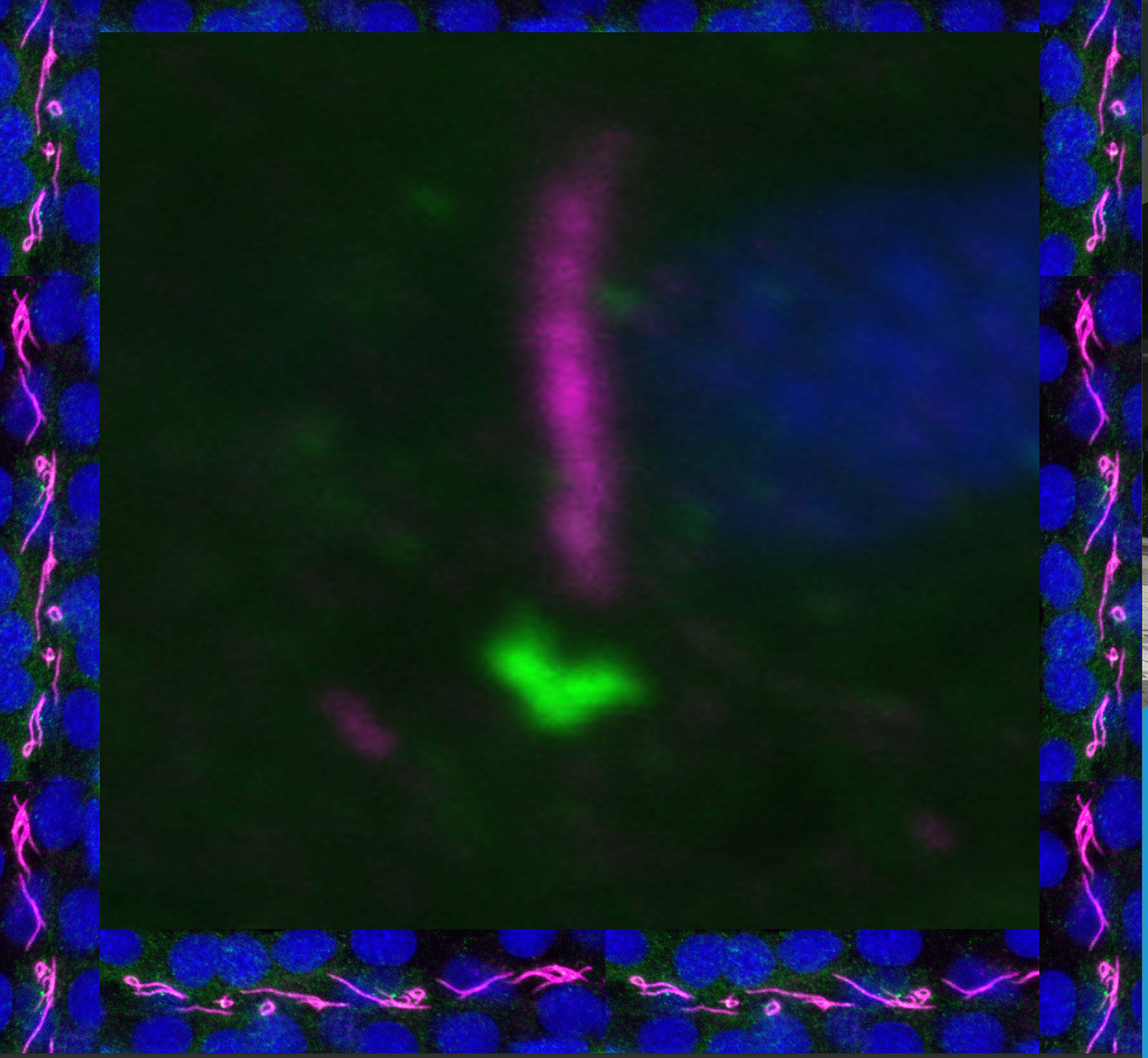 Confocal image showing localization of Odad1, a ciliary dynein docking complex member, at the base of cilia in the zebrafish left-right organizer. The border shows confocal image of cilia in the kidney duct where Odad1 localizes along the axonemes. Odad1 was detected using antibodies to GFP (fused to the C-terminus of Odad1) (green), while ciliary axonemes and nuclei were labelled with anti-acetylated tubulin antibodies (magenta) and DAPI (blue), respectively. See Research article by Lu et al. ( Development (2024) 151 (14): dev202737.). Image credit: Hao Lu and Sudipto Roy.