Oliver Dreesen

Dr Oliver Dreesen
Email: oliver_dreesen@asrl.a-star.edu.sg
Research themes: Skin Aging, Senescence, Premature Aging Syndromes, Nuclear lamina, Chromatin.
Senior Principal Investigator
Head of Cell Aging Laboratory
A*STAR Skin Research Laboratories, Singapore
Co-Principal Investigator, Adjunct Associate Professor
Mechanobiology Institute, National University of Singapore
Research themes: Skin Aging, Senescence, Premature Aging Syndromes, Nuclear lamina, Chromatin.
Biography
Dr. Oliver Dreesen is a Senior Principal Investigator and Head of the Laboratory for Cell Aging at the Agency for Science, Technology & Research (A*STAR) in Singapore. He served as President of the Skin Research Society (Singapore) from 2019-2021, was Chair of the Singapore Skin Research Conferences in 2019 and 2021; and is currently Treasurer of the Skin Research Society. He is a Co-Principal Investigator and Adjunct Associate Professor at the Mechanobiology Institute at the National University of Singapore. He received his Ph.D. from the Rockefeller University in New York City, USA.
Cell Aging Laboratory
The Cell Aging lab is 1) investigating fundamental processes of human aging, 2) characterizing novel biomarkers to detect senescent cells in vitro and in age-related skin pathologies in vivo, and 3) developing assays and tools to understand how senescence changes the function of cells, how they contribute to skin and organismal dysfunction and aging, and how to selectively eliminate or modulate their function (senolytics / senomorphics).
To achieve this, the lab focuses on elucidating the fundamental mechanism(s) of cell aging and senescence in Hutchinson-Gilford progeria (HGPS), a premature aging disorder that triggers early onset alopecia, aberrant skin pigmentation, skin fragility, lipodystrophy and skeletal abnormalities. Patients succumb to cardiovascular complication in their mid-teens. HGPS is caused by a dominant-negative mutant of the intermediate filament protein lamin A, termed progerin. The precise mechanism(s) how progerin impacts various aspects of cell physiology and causes disease remains unclear. The Dreesen lab pioneered the use of a doxycycline-inducible expression system, in conjunction with single-cell immunofluorescence microscopy, to delineate the temporal chain of events that occurs upon progerin expression and ultimately culminates in premature cell aging / senescence (Chojnowski et al., eLIFE 2015; Aguado et al., Nature Comm; 2019). The team found that progerin-induced heterochromatin loss already occurred in G0-arrested cells, whilst DNA damage occurred exclusively during late stages of DNA replication, and preferentially in cells with low levels of heterochromatin. Moreover, this experimental system allowed the team to determine which phenotypes would be reversible upon removal of the mutant protein (Chojnowski, Ong et al., Aging Cell 2020), and how farnesyltransferase inhibitors, currently the only FDA-approved treatment for HGPS patients, ameliorate the disease (Dreesen & Kennedy, Med 2021; Foo, Ong et al., Aging Cell 2024). Collectively, the results provide evidence for a mechanistic link between the nuclear envelope, chromatin organization, DNA replication and telomeres that is disrupted in progeria and other human diseases caused by perturbations of the nuclear envelope (Dreesen Biochem Soc Trans 2020).
Secondly, by studying cells from HGPS patients, Dr. Dreesen found that the nuclear periphery undergoes dramatic remodelling during cellular senescence. For instance, lamin B1, an intermediate filament protein expressed in all somatic cells, is downregulated in human skin cell types undergoing replicative senescence in vitro, as well as during chronological aging of human skin in vivo (Dreesen et al., J Cell Bio 2013, Nucleus 2013). By multiplexing skin cell type specific markers with lamin B1, the Dreesen group quantified the accumulation and clearance of senescent cells in different compartments of the skin after UV-exposure and upon regeneration, respectively (Wang et al., Sci Rep 2017). In collaboration with clinicians at Kyoto University, the team used senescence markers lamin B1 and HMGB1, in conjunction with proliferation markers, to identify and quantify senescent cell types in pre-cancerous actinic keratosis (AK) lesions (Wang et al., J Derm Sci 2022). Taken together, this novel biomarker facilitates the identification of senescent cells in age-related skin pathologies, including AK or solar lentigo (unpublished), upon exposure to environmental conditions such as pollution or UV-exposure, as well as before and after treatment with anti-aging compounds or drugs that selectively eliminate senescent cells, such as senolytics or senomorphics (Chin, Lee et al., Front in Phys 2023).
Thirdly, the team has established a number of experimental tools and assays to generate different senescent skin cell types to understand their function, and investigate how senescent cells impact their healthy neighbours in 2D and 3D reconstructed in vitro skin models (in collaboration with the Bonnard group).
Collectively, these studies have resulted in 44 peer-reviewed publications in leading journals including Nature, Journal of Cell Biology, eLIFE, Aging Cell, Nature Communications, Journal of Dermatological Science, Cell Stem Cell, iScience and Nucleus, as well a patent applications.

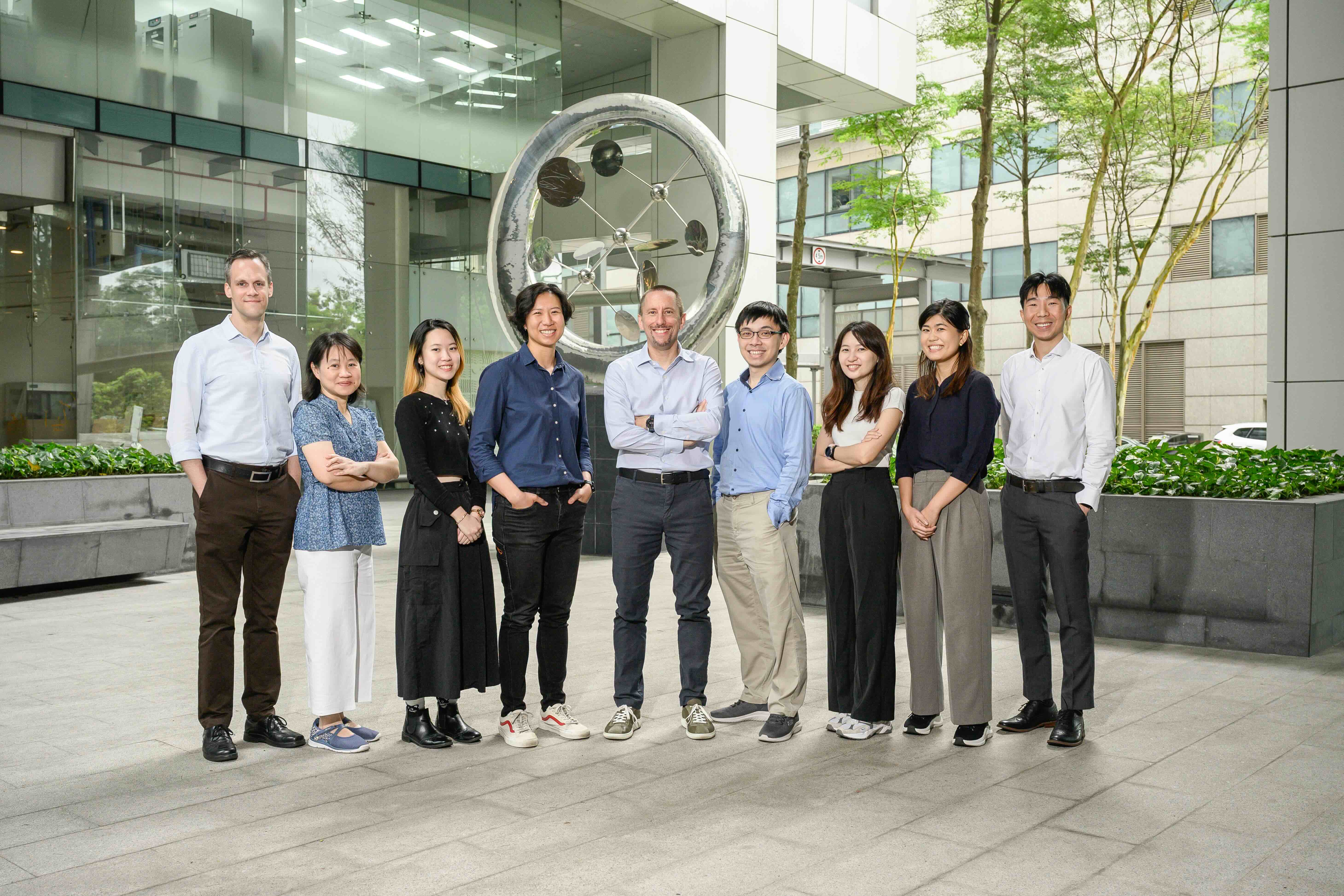
Dreesen's Lab Team
Left to Right: Ivo Johannes, Ong Peh Fern, Tristy Abigayle, Arnette Wong, Oliver Dreesen, Mattheus Foo, Lee Xin Er, Ng Pei Yi, Selwyn Low
Publications:
For an updated list of publications, please see the link below:
Foo XRM#, Ong PF#, Yap ZX, Maric M, Bong CJS, Droge P, Burke B, Dreesen O* (2024). Genetic and pharmacological modulation of lamin A farnesylation determines its function and turnover. Aging Cell, March 19; e14105. doi: 10.1111/acel.14105
Braun H, Xu Z, Chang F, Viceconte N, Rane G, Levin M, Lototska L, Roth F, Hillairet A, Sola AF, Khanchandani V, Dreesen O, Yang Y, Shi Y, Li F, Butter F, Kappei D (2023). ZNF524 directly interacts with telomeric DNA and supports telomere integrity. Nature Communications, Dec 12;14(1):8252. doi: 10.1038/s41467-023-43397-7.
Sotiropoulou K, Yumlu S, Hirano T, Maier M, Loh, A, Ong PF, Yim OS, Liu C, Vial E, Altunoğlu U, Nampoothiri S, de Silva D, Fischer-Zirnsak B, Hülya Kayserili H, Lai PS, Dreesen O, Kabashima K, Kornak U, Escande-Beillard N, Reversade B (2023). PYCR1 Levels Track with Premature and Chronological Skin Aging. MedRxiv May 28th doi.org/10.1101/2023.05.24.23289766
Jarrold BB, Tan CYR, Ho CY, Soon AL, Lam TT, Yang X, Nguyen C, Guo W, Chew YC, DeAngelis YM, Costello L, De Los Santos Gomez P, Przyborski S, Bellanger S, Dreesen O, Kimball AB, Oblong JE (2022). Early onset of senescence and imbalanced epidermal homeostasis across the decades in photoexposed human skin: Fingerprints of inflammaging. Exp Dermatol. Nov;31(11):1748-1760. doi: 10.1111/exd.14654.
Dainichi T, Nakano Y, Doi H, Nakajima S, Litman T, Narang V, Nakamizo S, Kawakami E, Guttman-Yasski E, Dreesen O, Reversade B, Kabashima K (2022). Epithelial stress-induced peptide (ESIP) is a potential master-regulator for an epithelial and protective response of the skin. Frontiers in Immunology, Feb 22,13:825032. doi:10.3389/fimmu.2022.825032
Wang AS, Nakamizo S, Ishida Y, Klassen G, Chong P, Wada A, Lim JSY, Wright GD, Kabashima K, Dreesen O* (2022). Identification and quantification of senescent cell types by lamin B1 and HMGB1 in Actinic keratosis lesions. Journal of Dermatological Science, Jan;105(1):61-64. doi10.1016/j.dermsci.2021.12.001
Tan CYR, Tan CL, Chin T, Morenc M, Ho CY, Rovito HA, Quek LS, Soon AL, Lim JSY, Dreesen O, Oblong JE, Bellanger S (2021). Nicotinamide prevents UVB- and oxidative stress-induced photoaging in human primary keratinocytes. Journal of Investigative Dermatology, Jun;142(6):1670-1681.e12. doi: 10.1016/j.jid.2021.10.021.
Giam M, Wong CK, Low JS, Sinelli M, Dreesen O, Rancati G (2020). p53 induces senescence in the unstable progeny of aneuploid cells. Cell Cycle, Dec;19(24):3508-3520. doi: 10.1080/15384101.2020.1850968.
Chojnowski A#, Ong PF#, Foo MXR, Liebl D, Hor LP, Stewart CL, Dreesen O* (2020). Heterochromatin loss as a determinant of progerin-induced DNA damage in Hutchinson-Gilford Progeria. Aging Cell, Mar;19(3):e13108. doi: 10.1111/acel.13108.
Aguado J, Sola-Cavajal A, Cancila V, Revechon G, Ong PF, Wallen Arzt E, Lattanzi G, Dreesen O, Tripodo C, Rossiello, Eriksson M, d’Adda di Fagagna F (2019). Sequence-specific inhibition of DNA damage response at telomeres improves the detrimental phenotypes of Hutchinson-Gilford Progeria Syndrome. Nature Communications, Nov 18;10(1):4990. doi: 10.1038/s41467-019-13018-3.
Ivanyi-Nagy R, Ahmed SM, Peter S, Dharshana Ramani P, Ong PF, Dreesen O, Dröge P (2018). The RNA interactome of human telomerase RNA reveals a coding-independent role for histone 1C mRNA in telomere homeostasis. eLIFE, Oct 25;7:e400037. doi: 10.7554/eLife.40037.
Chojnowski A, Sobota RM#, Ong PF#, Dreesen O, Stewart CL, Burke B (2018). 2C-BioID: an improved, two component BioID system that refines the identification of protein interactomes iScience, Dec 21;10:40-52. doi: 10.1016/j.isci.2018.11.023.
Szmyd R, Niska-Blakie J, Diril MK, Renck-Nunes P, Tzelepis K, Lacroix A, Van Hul N, Matos J, Dreesen O, Bisteau X, Kaldis P (2018). Premature activation of Cdk1 leads to mitotic events in S phase and embryonic lethality. Oncogene, Feb;38(7):998-1018. doi: 10.1038/s41388-018-0464-0.
Wang AS#, Ong PF#, Chojnowski A, Clavel C*, Dreesen O* (2017). Loss of lamin B1 is a biomarker to quantify cellular senescence in photoaged skin. Scientific Reports, Nov 15;7(1):15678. doi: 10.1038/s41598-017-15901-9.
Ling L, Camilleri ET, Helledie T, Samsonraj RM, Titmarsh DM, Chua RJ, Dreesen O, Dombrowski C, Rider DA, Galindo M, Lee I, Hong W, Hui JH, Nurcombe V, van Wijnen AJ, Cool SM (2016). Effect of heparin on the biological properties and molecular signature of human mesenchymal stem cells. Gene, Jan 15;576(1Pt 2):292-303. doi: 10.1016/j.gene.2015.10.039.
Chojnowski A, Ong PF, Wong ESM, Lim JSY, Mutalif RA, Navasankari R, Dutta B, Yang H, Liow YY, Sze SK, Boudier T, Wright GD, Colman A, Burke B, Stewart CL*, Dreesen O* (2015). Progerin reduces LAP2α:telomere association in Hutchinson-Gilford progeria syndrome. eLife, Aug 27;4;e07759. doi: 10.7554/eLife.07759.
Dreesen O*, Ong PF, Chojnowski A, Colman A* (2013). The contrasting roles of lamin B1 in cellular aging and human disease. Nucleus, Jul-Aug;4(4):283-90. doi: 10.4161/nucl.25808.
Dreesen O*, Chojnowski A, Ong PF, Zhao TY, Common JE, Lunny D, Lane EB, Lee SJ, Vardy LA, Stewart CL, Colman A* (2013). Consequences of Lamin B1 fluctuations on cellular proliferation and senescence. Journal of Cell Biology, Mar 4;200(5):605-17. doi: 10.1083/jcb.201206121.
Hovel-Miner G, Boothroyd CE, Mugnier M, Dreesen O, Cross GAM, Papavasiliou FN (2012). Telomere length affects the frequency and mechanism of antigenic variation in Trypanosoma brucei. PLoS Pathogens, 2012;8(8):e1002900. doi: 10.1371/journal.ppat.1002900.
Pomp O, Dreesen O, Leong D, Tan T, Meller-Pomp O, Colman A (2011). Unexpected X-chromosome skewing during culture and reprogramming of human somatic cells can be alleviated by exogenous telomerase. Cell Stem Cell, Aug 5;9(2):156-65. doi: 10.1016/j.stem.2011.06.004.
Boothroyd CE#, Dreesen O#, Leonova T, Ly KI, Figueiredo LM, Cross GAM, Papavasiliou FN (2009). A yeast-endonuclease-generated DNA break induces antigenic switching in Trypanosoma brucei. Nature, May 14;459(7244):278-81. doi: 10.1038/nature07982.
Dreesen O and Cross GAM (2008). Telomere length in Trypanosoma brucei. Experimental Parasitology, Jan;118(1):103-10. doi: 10.1016/j.exppara.2007.07.016.
Dreesen O and Cross GAM (2006). Consequences of telomere shortening on active VSG expression site in telomerase-deficient T. brucei. Eukaryotic Cell, Dec;5(12):2114-9. doi: 10.1128/EC.00059-06.
Dreesen O and Cross GAM (2006). Telomerase-independent stabilization of short telomeres in T. brucei. Molecular and Cellular Biology, Jul;26(13):4911-9. doi: 10.1128/MCB.00212-06.
Dreesen O, Li B and Cross GAM (2005). Telomere structure and shortening in telomerase-deficient Trypanosoma brucei. Nucleic Acids Research, Aug 9;33(14):4536-43. doi: 10.1093/nar/gki769.
Janzen CJ#, Lander F#, Dreesen O and Cross GAM (2004). Telomere length regulation and transcriptional silencing in KU80-deficient Trypanosoma brucei. Nucleic Acids Research, Dec15;32(22):6575-84. doi: 10.1093/nar/gkh991.
Kobayashi K, Ehrlich SD et al., (2003). Essential Bacillus subtilis genes. Proc. Natl. Acad. Sci. USA, Apr 15;100(8):4678-83. doi: 10.1073/pnas.0730515100.
Cruz Ramos H, Hoffmann T, Marino M, Nedjari H, Presecan–Siedel E, Dreesen O, Glaser P, Jahn D (2000). Fermentative metabolism of Bacillus subtilis: Physiology and regulation of gene expression. Journal of Bacteriology, Jun;182(11):3072-80. doi: 10.1128/JB.182.11.3072-3080.2000.
King N, Dreesen O, Stragier P, Pogliano K, Losick R (1999). Septation, dephosphorylation and activation of sigmaF during sporulation in Bacillus subtilis. Genes and Development, May 1;13(9):1156-67. doi: 10.1101/gad.13.9.1156.
Reviews & Meeting Reports:
Chin T, Lee XE, Ng PY, Lee Y, Dreesen O* (2023). The role of cellular senescence in skin aging and age-related skin pathologies. Frontiers in Physiology, Nov 22;14:1297637. doi: 10.3389/fphys.2023.1297637.
Dreesen O (2023). Dermatological Researchers in the World, The Nishinihon Journal of Dermatology, Volume 85 Issue 1, 67-67, https://doi.org/10.2336/nishinihonhifu.85.67
Ho CY and Dreesen O* (2021). Faces of Cellular Senescence in Skin Ageing. Mechanism of Ageing and Development, Sep;198:111525. doi: 10.1016/j.mad.2021.111525.
Dreesen O* (2020). Towards delineating the chain of events that cause premature senescence in the accelerated aging syndrome Hutchinson-Gilford progeria (HGPS). Biochemical Society Transactions, Jun 30;48(3):981-991. doi: 10.1042/BST20190882.
Foo XRM, Ong PF, Dreesen O* (2019). Premature aging syndromes: from patients to mechanism. Journal of Dermatological Science, Nov;96(2):58-65. doi: 10.1016/j.jdermsci.2019.10.003.
Nanba D* and Dreesen O* (2019). Meeting Report: Japan – Singapore International Skin Conference, Singapore April 10-12, 2019. Journal of Dermatological Science, 94, 253-257. doi:10.1016/j.jdermsci.2019.05.006
Wang AS and Dreesen O* (2018). Biomarkers of Cellular Senescence and Skin Aging. Frontiers in Genetics, Aug 23;9:247. doi: 10.3389/fgene.2018.00247.
Chojnowski A, Ong PF, Dreesen O* (2014). Nuclear lamina remodeling and its implications for human disease. Cell & Tissue Research, Jun;360(3):621-31. doi: 10.1007/s00441-014-2069-4.
Pomp O, Dreesen O, Colman A (2011). The ‘X-factor’ in cellular reprogramming and proliferation. Cell Cycle, Dec 1;10(23):3992-3. doi: 10.4161/cc.10.23.18253.
Dreesen O and Stewart CL (2011). Accelerated aging syndromes, are they relevant to normal human aging? Aging, Sept;3(9):889-95. doi: 10.18632/aging.100383.
Colman A* and Dreesen O* (2009). Pluripotent stem cells and disease modeling. Cell Stem Cell, Sept 4;5(3):244-7. doi: 10.1016/j.stem.2009.08.010.
Colman A and Dreesen O (2009). Induced pluripotent stem cells and the stability of the differentiated state EMBO Reports, Jul;10(7):714-21. doi: 10.1038/embor.2009.142.
Dreesen O and Brivanlou AH (2007). Signaling pathways in Cancer and Embryonic Stem Cells. Stem Cell Reviews, Jan;3(1):7-17. doi: 10.1007/s12015-007-0004-8.
Viewpoint & Opinion Articles:
Lee XE and Dreesen O* (2023). Sun exposure and its impact on keratinocyte senescence. Journal of Investigative Dermatology, Apr;143(4):529-530. doi: 10.1016/j.jid.2022.12.010
Dreesen O* and Kennedy BK (2021). Hutchinson-Gilford Progeria paves the way for novel targeted anti-aging therapies. Med (Cell Press), Apr 9;2(4):353-354. doi: 10.1016/j.medj.2021.03.005.
Dreesen O, Li B and Cross GAM (2007). Telomere structure and function in trypanosomes: a proposal. Nature Reviews Microbiology, Jan;5(1):70-5. doi: 10.1038/nrmicro1577.
Book Chapter:
Dreesen O* and Colman A* (2011). Book Chapter: Induced Pluripotent Stem Cells. Stem Cells: From Bench to Bedside. Second Edition.
A*STAR Open Access Repository
A*STAR celebrates International Women's Day

From groundbreaking discoveries to cutting-edge research, our researchers are empowering the next generation of female science, technology, engineering and mathematics (STEM) leaders.
Get inspired by our #WomeninSTEM
