
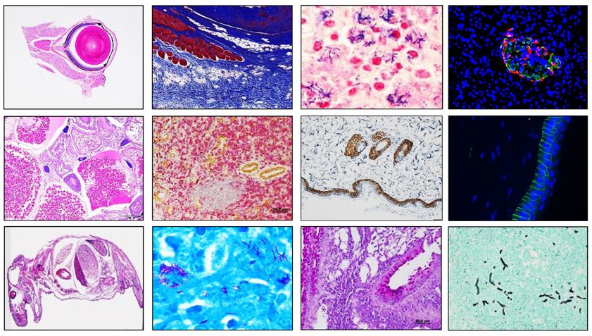
iCORE Pathology
A GLP-certified animal pathology laboratory house within A*STAR iCORE has been playing pivotal role in Singapore’s research ecosystems over 15 years. The laboratory provides research facilitation to A*STAR Research Institutes, Universities, Health sectors, Major pharmaceutical companies, and SMEs.
Staffed by highly experienced professionals and supported by robust infrastructures, the laboratory delivers valuable pathological insights that contribute to research projects, grant applications, and scientific publications across diverse disciplines. The laboratory was previously known as Advanced Molecular Pathology Laboratory (AMPL).
Key Services
- Animal Necropsy
- Simple and Complex Histo-Processing (Paraffin & Cryo)
- Embedding
- Sectioning
- H&E Staining
- Special Staining
- Immunohistochemistry (Brightfield & Immunofluorescence)
- Tissue Microarray
- Whole Slide Imaging
- Image Annotation and Image Analysis
- Clinical Chemistry Analysis
- Haematology Analysis
- Urine Analysis
- Animal Pathology Evaluation for Research Cases
- Animal Pathology Evaluation for Veterinary Diagnostics
- Clinical Pathology Evaluation for Research Cases (Human Tissues)
- Animal Pathology Toxicity Assessment
- Animal Pathology Toxicology Consultation
- GLP Training
Key Applications
Histology/Histopathology/clinical pathology supports for
Preclinical & basic research
- Phenotyping of animal models: characterize organs/tissues in knockout, transgenic, disease models
- Mechanism of disease studies: correlate microscopic changes with biochemistry/haematology
- Developmental biology: embryonic and postnatal tissue differentiation and defects
Drug discovery & development
- Safety pharmacology support: correlation of clinical signs with tissue lesions
- Efficacy assessment: histological evidence of treatment response or healing
- Toxicologic pathology : target organ toxicity, dose-response, Reversibility, MTD etc
- Biomarker validation: IHC, ISH, special stains to link structure and functional alterations
Gene, cell, and advanced therapies
- Tissue distribution studies: cell and gene therapy localization
- On-target/off-target effects: tissue-specific toxicity and inflammation
- Tumorigenicity assessments: cell therapy safety
Infectious disease research
- Pathogen localization by special stain, IHC.
- Host-pathogen interaction: inflammation, tissue damage patterns
- Vaccine studies: reaction at injection site, organ response
Device & biomaterial evaluation
- Implant biocompatibility
- Foreign body response, fibrosis, encapsulation
Methodological support
- Tissue processing, embedding, sectioning, special stains
- Immunohistochemistry & morphometry
- Digital pathology for quantitative image analysis
Key Equipment
- Leica Autostainer XL (Automated Slide Stainer)
- Leica CV5030 Coverslipper (Automated Slide Coverslipper)
- Leica CM3050 S Cryostat (Cryostat)
- Sakura Tissue-Tek TEC5 Embedding System (Paraffin Embedding System)
- Leica Bond-Max™ (Automated IHC stainer)
- Leica Bond-III™ (Automated IHC stainer)
- Roche Ventana Discovery Ultra System (Automated IHC stainer)
- Leica IPC Cassette Printer (Cassette Printer)
- Leica IPS Slide Printer (Slide Printer)
- Leica RM2265 (Microtome)
- Leica RM2235 (Microtome)
- Leica RM2255 (Microtome)
- Axioscan 7 Histological Slide Scanning System (Whole Slide Scanning System)
- Sakura Tissue-Tek VIP® 6 (Tissue Processor)
- Sakura Tissue-Tek VIP® 6 AI (Tissue Processor)
- Sysmex XN-1000 Automated Haematology Analyser (Haematology Analyser)
- Roche cobas® c 111 Clinical Chemistry Analyser (Clinical Chemistry Analyser)
- Uri-Trak 120 (Strip Analysis) Urine Analyser
- MTA-1 Beecher Tissue Arrayer (TMA)
- Olympus/Nikon Microscopes

Accreditations
Lab Certification:
- Good Laboratory Practice (GLP)
Board Certifications:
- ACVP (USA)
- DABT (USA)
- ERT (UK)
- RCPath (UK)
Collaboration and Authorship
If there are significant intellectual or scientific contribution by our staff, such as substantial contributions to conception and design, acquisition of data or analysis and interpretation of the data, co-authorship should be offered to the contributing staff and we will share responsibility and scientific accountability for the results.
Our People
The iCORE Pathology Platform is supported by a dedicated team who deliver consistent, end‑to‑end histopathology services. Partnering closely with researchers, the team provides timely, high‑quality tissue processing and analysis, ensuring dependable support across a broad range of research needs.
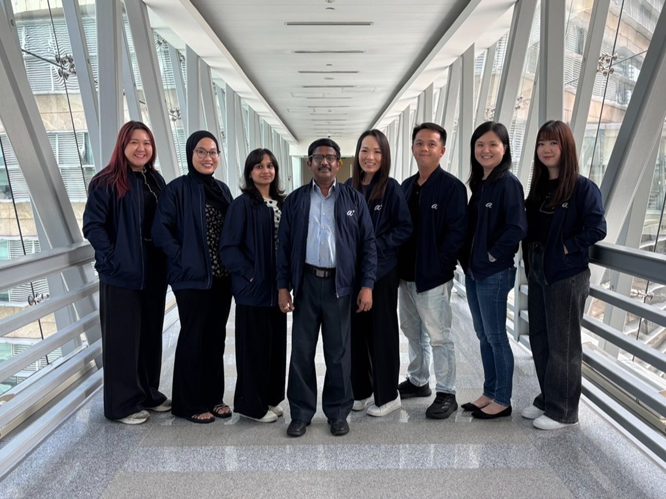
Team Members (from left to right): Maggie See; Atieqa Edora Eadi; Murugan Hemavarshini; Ravisankar Rajarethinam- Platform Lead; Ong Li-jin; Marcus Yong; Poh Suat Fang; Heng Shi Min
Our Documents
You may find the documents require for booking of our service here.
Histopathology Research Services
- iCORE Pathology Sample Request Form (Jul 26)
- Project Information for Pathology Evaluation (Jul 26)
- Antibody Receipt and IHC Test Questionnaire (Jul 26)
- Research Service List (Jul 26)
Animal Clinical Pathology
- Animal Clinical Pathology Sample Request Form (Jul 26)
- Animal Clinical Pathology Frequently Asked Questions (Jul 26)
Veterinary Diagnostics
- iCORE Pathology Vet Surgical Pathology Submission Form (Jul 26)
- Veterinary Surgical Pathology Services Pricelist (Jul 26)
- Vet Diagnostics Brochure (Jul 26)
Email us at iCOREPath@a-star.edu.sg to learn more about our platforms, services and how our team can support your research needs.
Telephone: 6586 9629