PI/Head: Shu Zhen CHONG, Ph.D.
Email: Chong_Shu_Zhen@a-star.edu.sg
Manager: Leonard TAN, Ph.D.
Email: Leonard_Tan@a-star.edu.sg
EXPLORE THE FULL POTENTIAL OF SPATIAL BIOLOGY BY COMBINING 3D LIGHT SHEET IMAGING WITH 2D MULTIPLEX ANALYSIS WITH THE SPATIAL BIOLOGY AND IMMUNE IMAGING PLATFORM
The A*STAR Singapore Immunology Network (SIgN), in partnership with Miltenyi Biotec, has established the Spatial Biology & Immune Imaging Centre of Excellence to advance next-generation biomedical research. The centre integrates innovative platforms that combine high-plex, spatially resolved molecular profiling with large-scale 3D visualization of intact tissues. This unique combination allows researchers to map proteins and gene expression within the native architecture of tissue while also capturing structural context across whole organs and biopsies. Supported by optimized preparation workflows and advanced analysis tools, the centre delivers reproducible, multiscale insights that connect molecular signatures with tissue- and organ-level organization, driving discoveries in immunology, oncology, neuroscience, and beyond.
In collaboration with


BACKGROUND
TECHNOLOGIES AND APPROACH
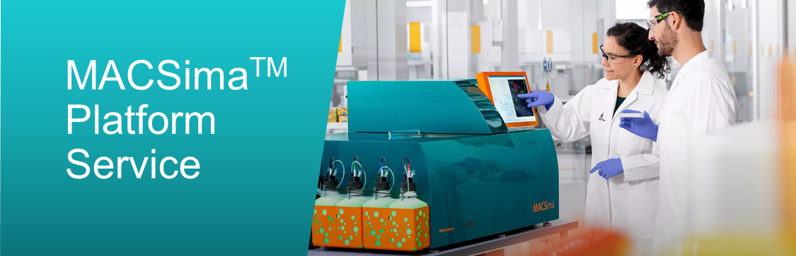
The Centre of Excellence boasts well-established protocols and expertise, offering comprehensive services spanning from sample preparation for image acquisition to thorough data analysis tailored to your requirements. Collaboratively, our SIgN Immune Imaging Platform, in conjunction with the Miltenyi Biotec team, will devise the optimal approach based on considerations such as sample size, quality, antibody panel, region-of-interest selection, and cost constraints. Additionally, we extend an Introductory package at a competitive rate to first-time users, facilitating exploration of the MACSimaTM Imaging System and optimizing sample and antibody panel designs.
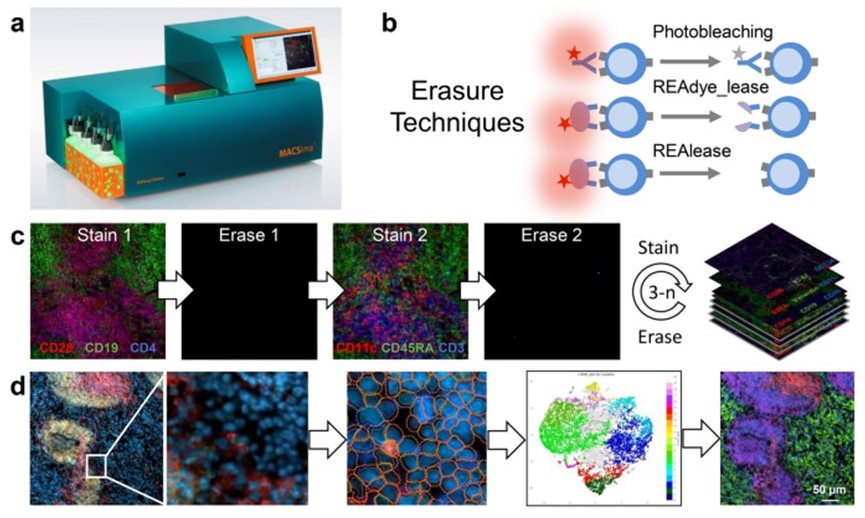
(Kinkhabwala et al, Nature, 2022)
Figure 1: Cyclic imaging utilizing the MACSimaTM Imaging System involves several key components. (a) The MACSimaTM System features fully automated robotic liquid handling and image acquisition capabilities. (b) Erasure techniques are employed, which may include photobleaching of the dye or disruption of the labeling conjugate using release reagent. The release reagent induces rapid detachment of the fluorescent dye alone (REAdye_lease Antibodies) or disrupts the labeling conjugate, resulting in spontaneous dissociation of the monomerized antibody fragments and the fluorescent dye (REAlease Antibodies) from their target epitopes. (c) Cyclic imaging involves repeated imaging cycles. (d) Image analysis is conducted using the MACS iQ View Software, which includes cellular segmentation, clustering, and visualization of clustered cells across the original image.
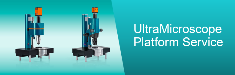
The UltraMicroscope platform is a high-performance light-sheet fluorescence microscopy system designed for detailed 3D imaging of optically cleared biological samples. The latest UltraMicroscope Blaze™ advances this capability with full automation, including autofocus, objective exchange, sample handling, and chromatic correction, and a LightSpeed mode that enables imaging up to 60 times faster than before, such as capturing an entire mouse brain in under three minutes. These features make it ideal for high-throughput, reproducible imaging across diverse specimens, from whole rodent organs to human biopsies and organoids.
To complement hardware, integrated MACS® iQ View software provides a seamless analysis pipeline with stitching, deconvolution, denoising, and batch processing optimized for light-sheet datasets. At SIgN Immune Imaging platform, we operated in collaboration with Miltenyi Biotec and the A*STAR Microscopy Platform (AMP), supported by more than a decade of expertise in tissue clearing and imaging. Optimized workflows cover a wide range of tissue types, including brain, lung, liver, skin, and tumors, and services span full sample processing, imaging, and introductory support packages for first-time users. Together, this pipeline delivers robust and reproducible 3D insights to accelerate research in immunology, oncology, neuroscience, and beyond.
OTHER PLATFORM EQUIPMENT LIST (OPEN ACCESS)
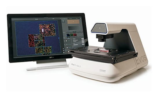
1. EVOS FL Auto 2 Cell Imaging System (Invitrogen)
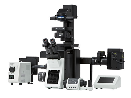
2. IX-83 Slide Scanner (Olympus)
3. CM1950 Cryostat (Leica)