
Florian WINAU
Biography
Dr Florian Winau is an immunologist whose research focuses on lymphocyte biology, antigen presentation and the role of lipids in immune responses. He studied medicine in Berlin, Paris and London, and trained as an MD before pursuing a research career in basic science. He completed residency duties as an assistant doctor in Internal Medicine at Charité Hospital in Berlin, before undertaking postdoctoral training in the Department of Immunology at the Max Planck Institute for Infection Biology in Berlin. During his postdoc time, he studied lipid antigen presentation by CD1 molecules, including lipid loading by saposins, and cross-presentation to CD8 T cells.
Dr Winau later established his laboratory at the Immune Disease Institute in Boston, which subsequently merged with Boston Children’s Hospital. His research expanded to activation mechanisms of innate-like T lymphocytes such as NKT cells, the role of CD1a-expressing Langerhans cells in skin inflammation, and the impact of lipid scramblase on T cell exhaustion. Recent work from his group has uncovered mechanisms in liver fibrosis, prosaposin-mediated tumour immunity, and the role of the lipid Gb3 in B cell and antibody responses. At Harvard Medical School, Dr Winau was a professor in the Departments of Microbiology, Immunology and Pediatrics. His work has been supported by NIH and industry grants and recognised through awards including the Robert Koch Prize for Immunology (Postdoctoral Fellows), a Smith Family Award for Excellence in Biomedical Research, and an Endowed Investigatorship of the Program in Cellular and Molecular Medicine.
Main Appointment
- Senior Principal Scientist III, Singapore Immunology Network (SIgN, A*STAR, Singapore)
Research Focus
The Winau Lab studies the pathophysiology of the immune system, focusing on how diverse immune cell types function and how these functions can go awry in disease. The lab seeks to decipher immune phenotypes in vivo, uncover the underlying molecular mechanisms, and translate mechanistic insights into therapeutic targets.
The lab’s approach integrates functional immunology, molecular mechanism and translation to therapy. Building on expertise in antigen presentation, T cell biology and lipid immunology, the lab investigates B lymphocyte functions and humoral immunity in viral infections, tumour immunology, organ-associated diseases such as fibrosis of the liver and lung, and inflammatory pathologies of diverse tissues, including the skin. This reflects a holistic approach to immunology, spanning innate and adaptive lymphocytes to uncover disease mechanisms and identify future therapeutic opportunities.
Key Research Themes
I. Lipid regulation of antibody responses
Recent work from the lab identified a vital function of the lipid globotriaosylceramide (Gb3) in regulating the affinity and diversity of antibody responses (Science, 2024). Gb3 is expressed on germinal centre B cells and binds the signalling hub protein CD19, facilitating B cell receptor downstream activation. The lab also showed that Gb3 can act as an immune potentiator in influenza vaccination, promoting broadly neutralising antibodies and cross-protection (Science, 2024).
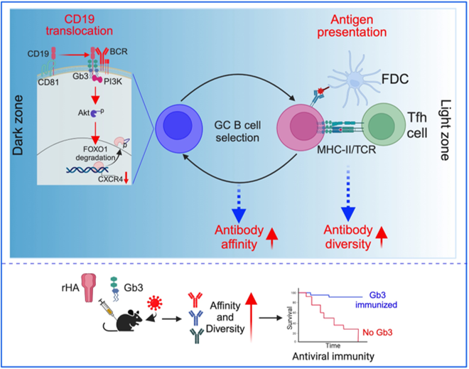
Figure 1. Gb3 drives affinity and diversity of antibody responses and protects against viral infection. Gb3 binds CD19 in germinal centre B cells to support B cell receptor signalling and antibody affinity maturation. It also regulates MHC-II levels, supporting broader antibody responses and protection during influenza vaccination.
II. Saposins and tumour antigen processing
The lab has shown that saposins, a family of lipid-interacting proteins, are critical for the processing of membrane-associated tumour antigens and the promotion of cancer immunity (Science, 2024). Tumours can induce hyperglycosylation of prosaposin (pSAP), leading to immune evasion by impairing antigen processing and inhibiting tumour-specific T cell responses. Targeting recombinant pSAP to dendritic cells restored antigen processing and facilitated protection against cancer (Science, 2024).
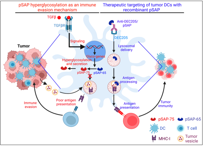
Figure 2. Mechanism of prosaposin-based immune escape and its targeting for immunotherapy of cancer. TGF-β induces hyperglycosylation and secretion of prosaposin in tumour dendritic cells, reducing saposin availability in the endolysosomal compartment and impairing antigen processing and presentation. Targeting recombinant prosaposin to tumour dendritic cells via DEC205 restores antigen processing and presentation, reinvigorates T cell responses and promotes tumour immunity.
III. Fibrosis according to GARP
The lab’s studies on hepatic stellate cells, a lipid-storing stromal cell type in the liver, revealed that these cells highly express GARP, a docking molecule for latent TGF-β. Surface-expressed GARP was shown to be essential for TGF-β activation by stellate cells, and ablation of GARP specifically in stellate cells reduced liver fibrosis (J. of Hepatology, 2023). This work identifies GARP as a potential target for antifibrotic therapy, with possible relevance to fibrotic diseases affecting the liver, kidney and lung.
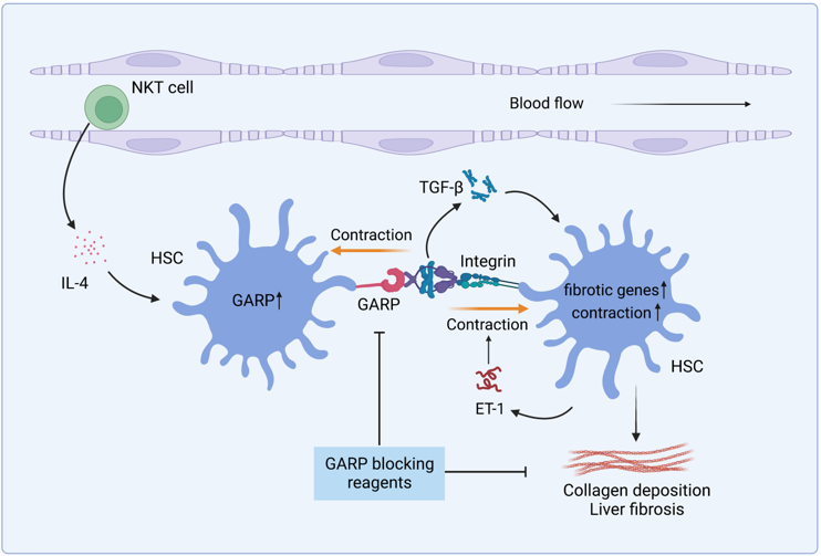
Figure 3. Mechanism of GARP-mediated liver fibrosis. Cytokines like IL-4 produced by NKT cells induce GARP expression on activated hepatic stellate cells (HSCs). GARP binds to latent TGF-β together with integrins, and with the help of cell contraction, releases the mature TGF-β. Activated TGF-β works on HSCs to promote fibrotic gene expression and endothelin-1 (ET-1)-mediated cell contraction, which leads to collagen accumulation and liver fibrosis.
IV. CD1 molecules in inflammation
Previous work from the lab showed that CD1a-expressing Langerhans cells trigger skin inflammation (Nature Immunology, 2016). Unlike classical MHC-I molecules, CD1 molecules present lipid antigens to T cells, and CD1a can present both exogenous and self-lipids to T lymphocytes. These findings established CD1a-mediated T cell activation as an important mechanism in inflammatory skin diseases and are now part of textbook knowledge (Paul’s Fundamental Immunology, 2021). Based on these findings, the lab developed a blocking antibody against CD1a that entered clinical trials for skin inflammation, including atopic dermatitis.
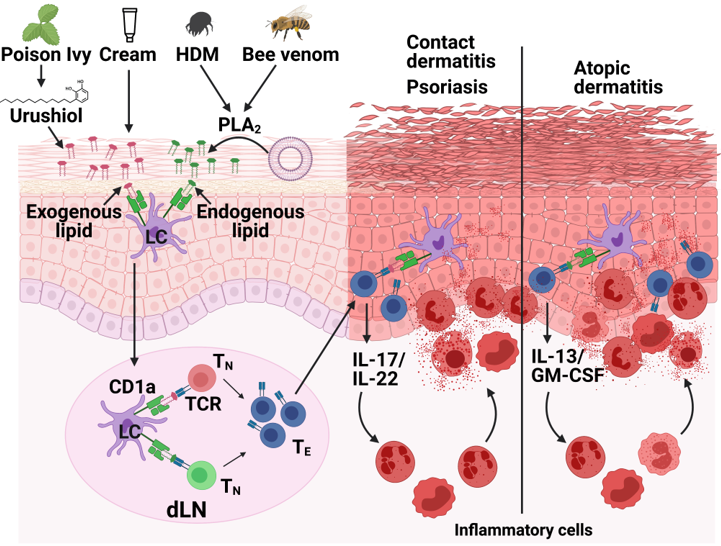
Figure 4. The role of CD1a on Langerhans cells in skin inflammation. External triggers can lead to lipid antigens being captured by Langerhans cells (LCs) in the skin. Activated LCs migrate to draining lymph nodes to present lipid antigens via CD1a, priming CD1a-restricted T cells. These activated lymphocytes return to the skin and deploy effector functions, contributing to skin inflammation. Depending on the trigger and immune response, CD1a-mediated T cell activation is important in various skin diseases.
HDM = house dust mite; PLA2 = phospholipase A2; TN = naïve T cell; TE = effector T cell; dLN = draining lymph node.
Lab Members
| Postdocs (Ph.D) |
|---|
| Tony Chu |
| May Yin Lee |