GIS RESEARCHERS DEVELOP FISHNCHIPS TO VISUALIZE DIFFERENT CELL TYPES WITHIN A DENSELY PACKED TISSUE
15 March 2024 - In biology, cells are the fundamental units of life, playing vital specialized roles like neurons transmitting signals or immune cells fighting disease. Traditionally, the goal of spatial transcriptomics has been to identify individual cell types using individual marker genes within a tissue sample. But this can be as difficult as spotting a particular person from a distance in a crowd. Just as individual features may be indistinguishable from afar, markers for certain cell types can also be too faint or blurry.
A team of researchers from A*STAR’s Genome Institute of Singapore (GIS) recently published a paper in Nature Communications to address this challenge. Principal Investigator and Group Leader at GIS’ Laboratory of Imagenomics, Dr Kok Hao Chen, Dr Nigel Chou and their team are early developers of spatial transcriptomics technologies in their research and wanted to derive an assay that would be more cost-effective and scalable. By using sets of genes, they developed FISHnCHIPs (Fluorescence In Situ Hybridization of Cellular HeterogeneIty and gene expression Programs), which maintains or even improves the robustness and sensitivity of the assay. The FISHnCHIPS method elegantly solves this challenge by looking at combinations of co-expressed markers instead of individual markers. The key insight is that genes involved in the same biological processes or pathways are co-localized within the same cells. Rather than relying on individual marker genes, FISHnCHIPs targets the entire gene programs. This allows it to rely on the multidimensional molecular "fingerprints" to robustly detect and distinguish different cell types. This enhanced sensitivity allows rapid, sensitive, and robust whole-tissue profiling under low magnification, and applicable for analyses of clinical tissue samples with low RNA quality. Remarkably, FISHnCHIPs also works robustly in frozen human samples, opening new avenues for discovering the spatial origin of rare cell types in disease. For more information, read the full article at: Protocols & Methods
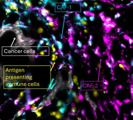
The colon tumor microenvironment imaged with FISHnCHIPs, showing two major types of fibroblasts cells (CAF) and their interactions with the immune and cancer cells.
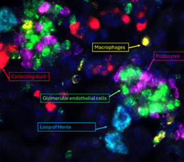
Kidney tissue imaged with FISHnCHIPs, showing cell types in and surrounding the Bowman’s capsule.