The global shortage of pathologists and rising cancer incidences have strained healthcare resources, leading to delayed and inadequate patient care. Our AI-based digital pathology (AIDP) platform addresses these challenges by providing fast, accurate, and accessible diagnostic tools. This platform aims to overcome ecosystem data and infrastructure issues, lack of production pipelines, and poor scalability of AI diagnostic models across different sites. By establishing an AI-powered diagnostic platform and developing necessary intellectual properties like A!HistoClouds and A!MagQC as demonstrated in panel A, we support AI model optimization and generalization. Additionally, creating a national annotated DP database and standardized annotation schemes enhances the development and deployment of AI solutions in clinical settings.
Our AIDP program’s goals include restoring image quality for cytology/pathology samples, developing a framework for AI models such as A!Prostate for Gleason grading, and utilizing generative AI to reduce dependence on human annotations. We also plan to implement autonomous cycled reinforcement learning to improve AI model optimization and generalization. By validating clinical AI pathological diagnostic models through international deployment and local cross-validation, we ensure high performance and accuracy. Our collaboration with institutions like A*STAR, NUH, SGH, and TTSH allows us to develop a comprehensive digital pathology development and adoption platform, integrating AI with a real-world pathological database. This initiative promises to enhance healthcare efficiency, generate commercially appealing intellectual properties, and stimulate job creation and talent development, positioning Singapore as a leading hub for AI-based diagnostics.
Establishing a Comprehensive Pipeline for Medical-Class Digital Pathology Diagnostic Models
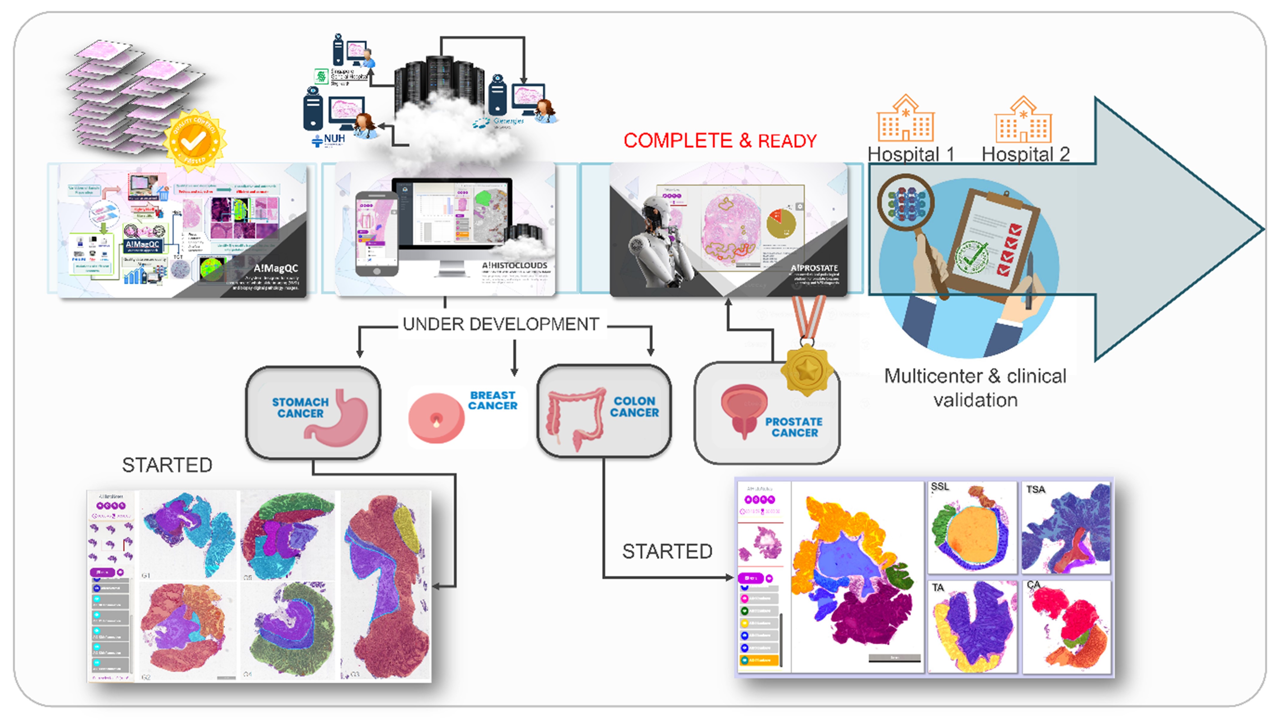
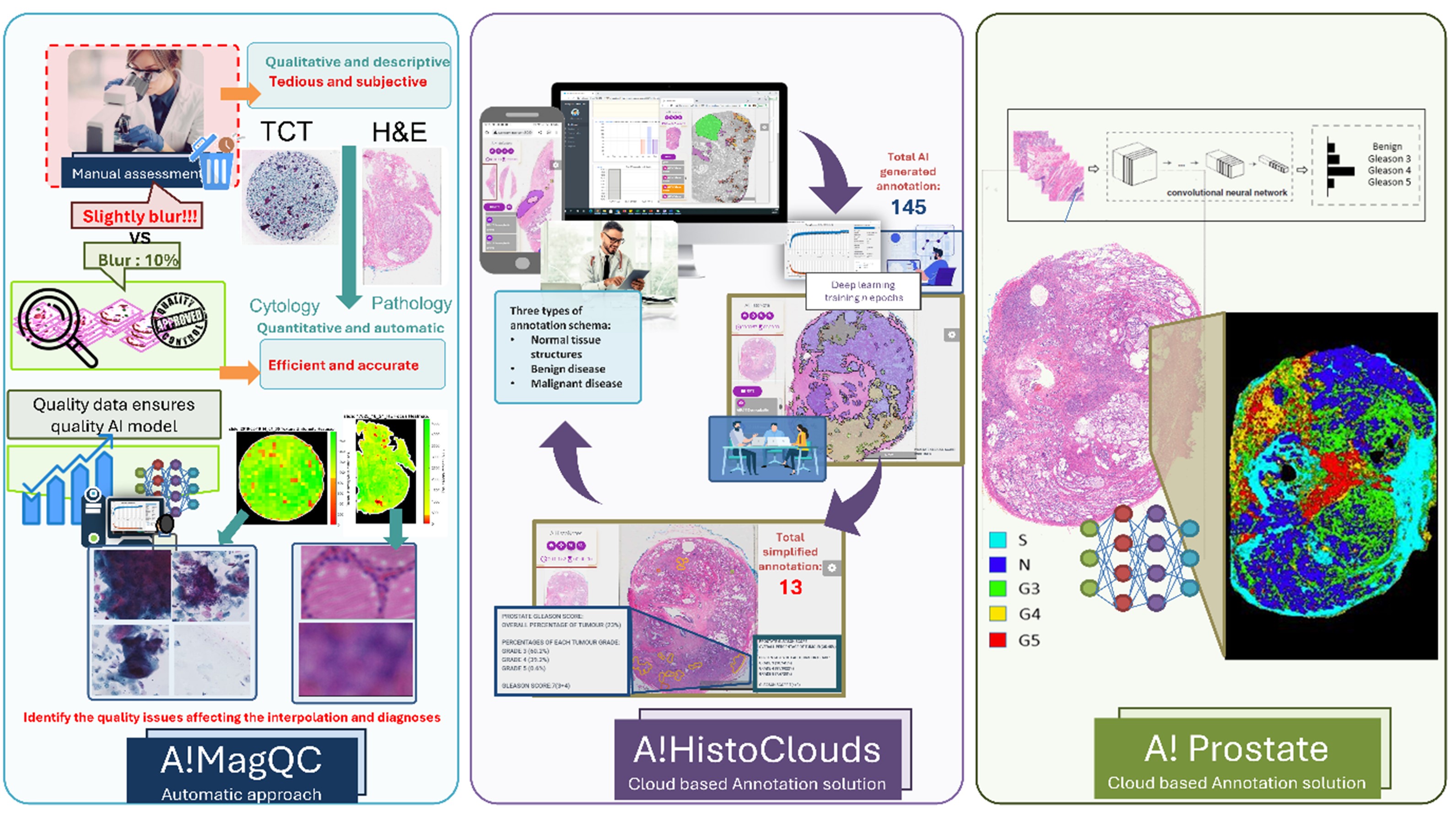
The establishment of a comprehensive pathological AIDP model development framework is essential to address the current gaps in standardized AI development pipelines across various diseases. Our innovative framework integrates general AI model optimization, generalization, and validation processes, supported by advanced tools like A!HistoClouds and A!MagQC. This pipeline, spanning from sample preparation to clinical decision-making, includes standard operating protocols (SOPs) for quality control and optimized imaging solutions. Large-scale DP data annotation and server/cloud-based storage bridge the gap between biomedical research and AI fields. Quality control (QC) and generalization are pivotal in this framework. A!MagQC ensures the consistency and reliability of image quality, transforming traditional subjective methods into objective, automated processes. This tool quantitatively assesses common image quality issues, ensuring high-quality data for AI model training and application. A!HistoClouds, on the other hand, facilitates pathologist-AI interaction (PAI), tele-pathology, and assistive pathological diagnosis, accelerating data curation through faster semi-automatic annotation. The development of medical-class R&D pipelines, workflows, and iterative optimization protocols from local and international data flows enhances the generalizability and robustness of AI models, ensuring their applicability across diverse clinical settings. By integrating these advanced QC and generalization methods, our framework aims to produce reliable, efficient, and scalable AI diagnostic solutions, significantly improving healthcare outcomes.
Revolutionizing Quality Control in AI Digital Pathology: The A!MagQC System
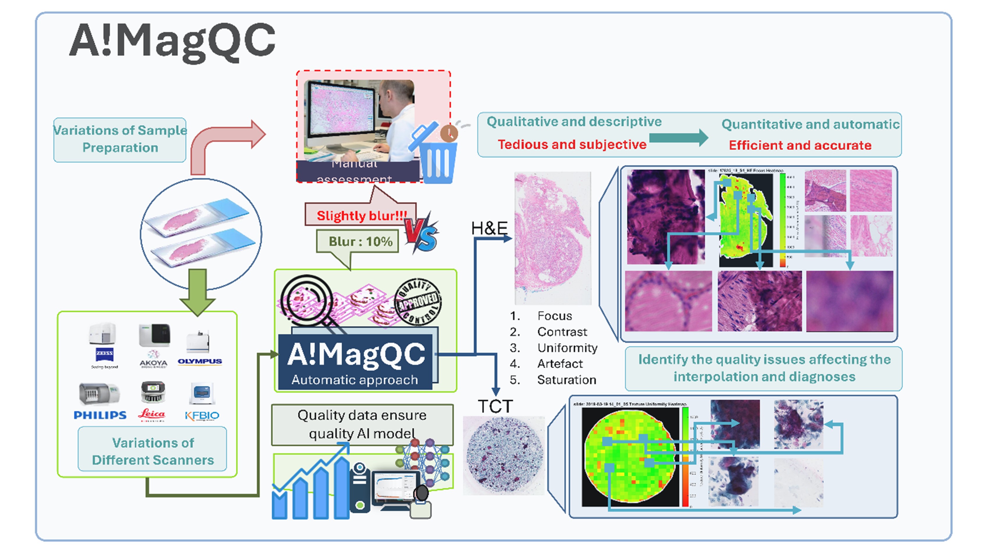
We designed A!MagQC to revolutionize image quality assessment, transforming traditional subjective, manual, and qualitative glass-slide QC into an objective, automated, and quantitative system. By applying advanced image processing techniques, A!MagQC comprehensively evaluates digital pathology whole slide images (WSI) across five critical categories: I) Out of focus, II) Contrast, III) Saturation, IV) Texture uniformity, and V) Artifacts. An image patch is deemed “low quality” if A!MagQC detects two or more issues among these categories, subsequently assigning a quantitative score to determine the overall quality of each WSI. A!MagQC has demonstrated remarkable efficiency, significantly reducing annotation costs and manpower effort while ensuring optimal scanner performance. In our program, A!MagQC will serve as the indispensable “gate-keeper” for our large-scale, high-quality annotated database, underscoring the pivotal role of quality control in AI digital pathology.
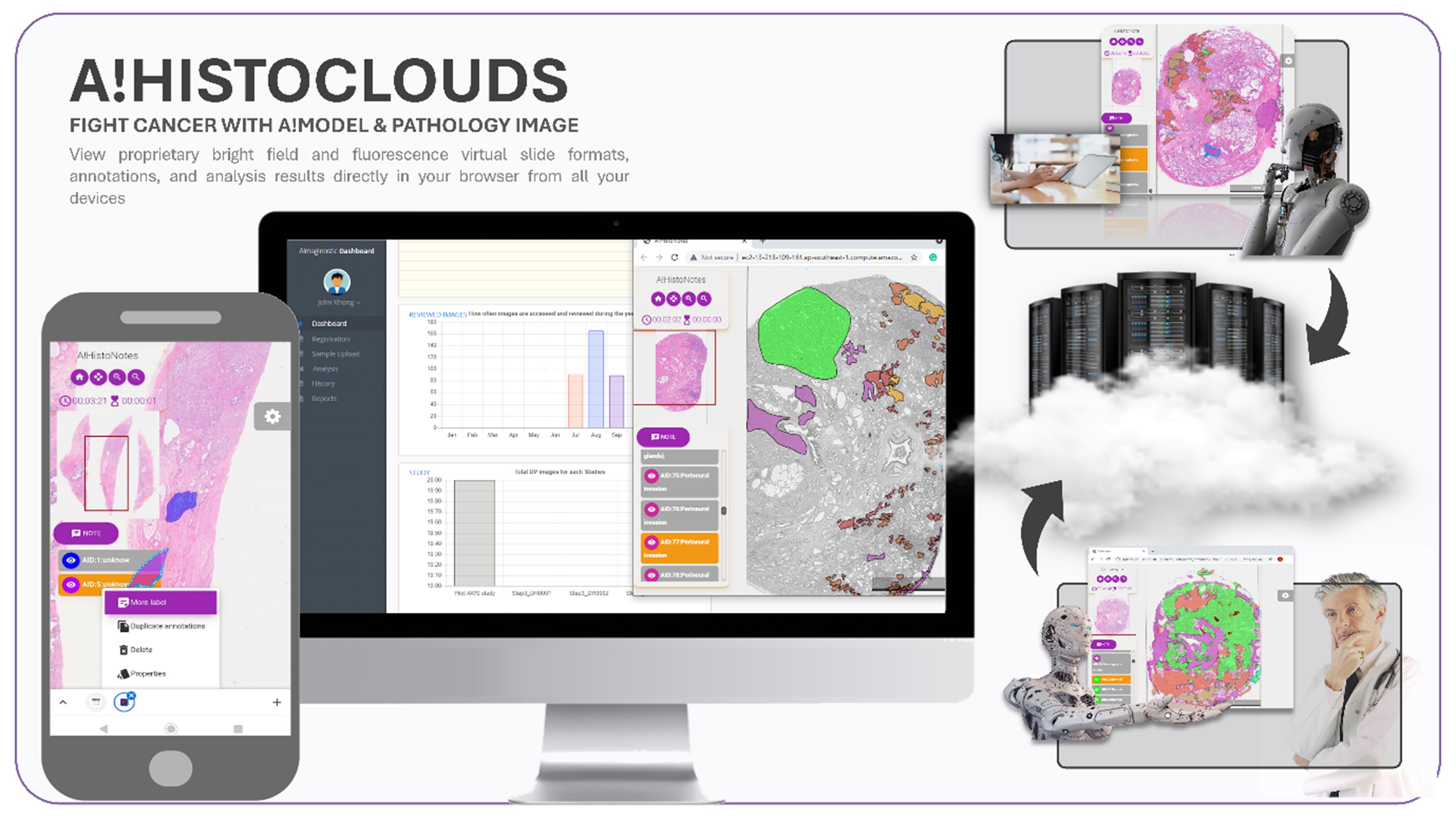
A!HistoClouds: Revolutionizing AI-Driven Pathological Annotation
We have developed A!HistoClouds, a user-friendly image annotation platform that integrates AI capabilities and Pathologist-AI interaction (PAI), enabling efficient online annotation and tele-pathology. As a cloud-based and local server-compatible solution, A!HistoClouds allows pathologists to work anytime, anywhere, and facilitates seamless collaboration. A!HistoClouds enhances the concept of PAI by bridging pathologists’ expertise and AI, serving as a key component in the evolution of online AI models. Demonstrated in the workflow, PAI refines AI models based on pathologists’ annotations. The platform supports well-defined annotation schemes tailored to different diseases, adhering to WHO/Association standards. Its AI model-agnostic design ensures compatibility with various AI diagnostic models, providing a holistic integration. Built with a human-centric approach, A!HistoClouds post-processes AI output into consumable visual information, streamlining the diagnostic process. The platform offers a highly interactive viewer, enabling pathologists to navigate large digital pathology images (gigapixel) smoothly. Using a pyramid image approach, similar to Google Maps, pathologists can dynamically view, zoom, and draw annotations on pathological images via the A!HistoClouds WSI viewer, enhancing both usability and efficiency.
Enhancing AI Diagnostic Accuracy Through Adaptive Generalization Techniques
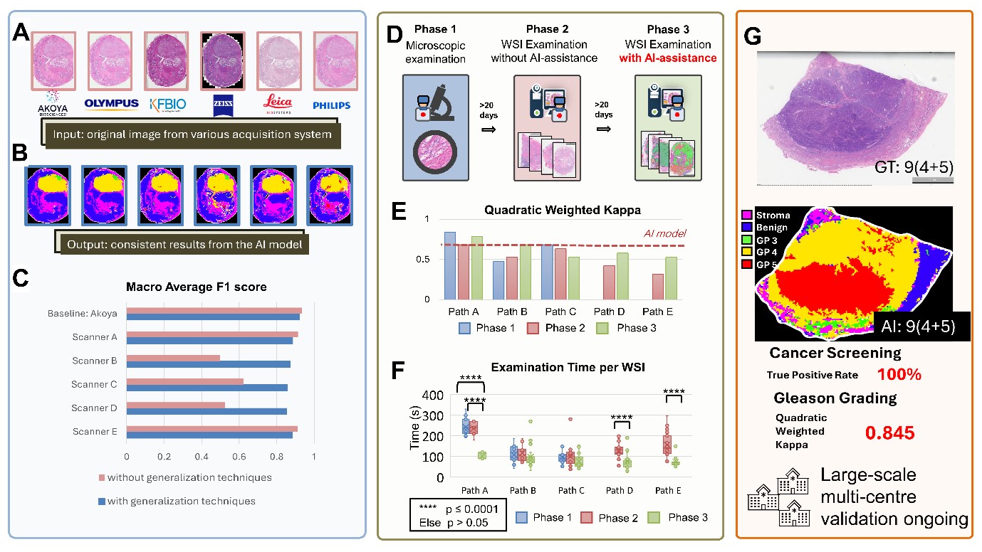
Development and training of our AI-enhanced solution began when scanned images were uploaded to A!HistoClouds, where clinical pathologists annotated regions of interest (ROIs) and assigned labels to them (stroma, normal glands, Gleason Pattern 3-5, for example in prostate diagnosis). After cropping the images into small patches and splitting the data into training and testing sets, we trained a convolutional neural network (CNN) using this training dataset to classify image patches into different label categories. On a per-image patch level, the sensitivity, specificity, positive predictive value, and negative predictive value were 93.0%, 92.7%, 96.3%, and 81.5%, respectively. We then visualized the prediction results on whole slide level by applying the model to the entire image. Comparing the generated heat maps and ground truth (pathologists’ annotations and Gleason scores), the predictions made by the AI model were well-matched with the pathologists’ diagnoses. For every different scanner brand, the digitization techniques and parameters are different. Therefore, the images of a single glass slide scanned by different scanners have varied appearances. To overcome this variation issue, we developed an adaptive solution by applying generalization techniques, named as Image Appearance Migration (IAM) to generate consistent results. Our strategy was to standardize images across these scanners and reduce image inconsistencies. Additionally, we further enhanced the generalizability of the models by enlarging the original training dataset using colour augmentation to simulate the appearance variations of different scanners during training. After applying generalization techniques, our AI models demonstrated increased accuracy and consistency among different scanners, potentially other variation in the upstream steps such as staining. Our experimental results indicate that our model is independent of the scanner hardware used, demonstrating its generalization ability across different scanning devices.
| Principal Investigator | YU Weimiao | [View Bio] |
| Senior Scientist | ONG Kok Haur |
| Scientist | YUAN Chengxiang |
| Scientist | LU Haoda |
| Scientist | SIMONGE Piumi |
| Lead Research Officer | HAO Han |
| Senior Research Officer | HUO Xinmi |
| Senior Research Officer | LI Longjie |
| Research Officer | JIANG Yijing |
| Research Officer | TAN Jun Aun |
| Research Officer | MONZON Eric |
| PhD Student | YOO Sehwan |
Xinmi Huo#, Kok Haur Ong#, Kah Weng Lau, Laurent Gole, Char Loo Tan, Chongchong Zhang, Yonghui Zhang, Xiaohui Zhu, Longjie Li, Hao Han, David Young, Haoda Lu, Jun Xu, Wanyuan Chen, Stephan J Sanders, Lee Hwee Kuan, Susan Swee-Shan Hue&, Weimiao YU&, Soo Yong Tan&. Comprehensive AI Model Development for Gleason Grading: From Scanning, Cloud-Based Annotation to Pathologist-AI Interaction. (2024). Nature - Communication Medicine. (IF NA)
Chunyue FENG#, Kokhaur ONG#, David M. YOUNG#, Bingxian CHEN, Longjie LI, Weizhong GU, Fei LIU, Hongfeng TANG, Manli ZHAO, Min YANG, Kun ZHU, Limin HUANG, Qiang WANG, Xinmi HUO, Gabriel P.L. MARINI, Haoda LU, Kun GUI, Hao HAN, Stephan J. SANDERS, Lin LI&, Weimiao YU&, and Jianhua MAO&. Artificial intelligence-assisted quantification and assessment of whole slide images for paediatric kidney disease diagnosis. (2024). Bioinformatics. (IF = 6.9)
Emanuela Frittoli#, Andrea Palamidessi#, Fabio Iannelli, Federica Zanardi, Stefano Villa, Leonardo Barzaghi, Hind Ando, Valeria Cancila, Galiba Beznuskenko, Giulia Della Chiara, Massimiliano Pagani, Chiara Malinverno, Dipanjan Bhattacharya, Federica Pisati, Weimiao Yu, Viviana Galimberti, Giuseppina Bonizzi, Emanuele Martini, Alexander Mironov, Chiara Rossi, Giovanni Bertalot, Marco Lucioni, Cristiuano Perini, FRancesco Ferrari, Chiara Lanzuolo, Guilherme Nader, Marco Foiani, Matthieu Piel, Roberto Cerbino, Fabio Giavazzi&, Claudio Tripodo&, Giorgio Scita&. Tissue fluidification promotes a cGAS/STING-mediated cytosolic DNA response in invasive breast cancer. (2023). Nature Material. (IF = 41.2)
Longjie Li#, Arun Mouli Kolinjivadi#, Kok Haur Ong, David M Young, Gabriel Pik Liang Marini, Sock Hoai Chan, Siao Ting Chong, Ee Ling Chew, Haoda Lu, Laurent Gole&, Weimiao Yu&, Joanne Ngeow&. Automatic DNA replication tract measurement to assess replication and repair dynamics at the single-molecule level. (2022). Bioinformatics. (IF = 6.9)
Xiaohui Zhu#, Xiaoming Li#, Kokhaur Ong#, Wenli Zhang, Wencai Li, Longjie Li, David Young, Yongjian Su, Bin Shang, Linggan Peng, Wei Xiong, Yunke Liu, Wenting Liao, Jingjing Xu, Feifei Wang, Qing Liao, Shengnan Li, Minmin Liao, Yu Li, Linshang Rao, Jinquan Lin, Jianyuan Shi, Zejun You, Wenlong Zhong, Xinrong Liang, Hao Han, Yan Zhang, Na Tang, Aixia Hu, Hongyi Gao, Zhiqiang Cheng, Li Liang&, Weimiao Yu&, Yanqing Ding&. Hybrid AI-assistive diagnostic model permits rapid TBS classification of cervical liquid-based thin-layer cell smears. (2021). Nature Communication. (IF = 16.6)
Laurent GOLE#, Joe YEONG#, Jeffery Chun Tatt LIM, Kok Haur ONG, Aye Aye THIKE, Yong Cheng POH, Sidney YEE, Jabed Iqbal, Bernett LEE*, Wanjin HONG&, Weimiao YU& and Puay Hoon TAN&. Quantitative stain-free imaging and digital profiling of collagen structure reveal diverse survival of triple negative breast cancer patients. (2020), Breast Cancer Research. (IF = 5.9)
Shyi-Chyi Wang, Tricia Yu Feng Low, Yukako Nishimura, Laurent Christophe Michel Gole, Weimiao Yu&, Fumio Motegi&, Cortical forces and CDC-42 control clustering of PAR proteins for C. elegans embryonic polarization. (2017). Nature Cell Biology. (IF = 28.2)
Chiara Malinverno, Salvatore Corallino, Fabio Giavazzi, Martin Bergert, Qingsen Li, Marco Leoni, Andrea Disanza, Emanuela Frittoli, Amanda Oldani, Emanuele Martini, Tobias Lendenmann, Gianluca Deflorian, Galina V. Beznoussenko, Dimos Poulikakos, ONG Kok Haur, Marina Uroz, Xavier Trepat, Dario Parazzoli, Paolo Maiuri, Weimiao Yu, Aldo Ferrari, Roßberto Cerbino, Giorgio Scita. Endocytic re-awakening of motility in jammed epithelia. (2017) Nature Materials. (IF = 41.2)
Adam Cliffe#, David P. Doupé#, HsinHo SUNG, Isaac Kok Hwee LIM, Kok Haur ONG, Li CHENG, Weimiao YU&. Quantitative analysis of complex individual cell behaviors in highly coordinated in vivo collective cell migration. (2017). Nature Communication. (IF = 16.6)
 | YU WeiMiao Principal Scientist Email: yu_weimiao@a-star.edu.sg Research Group: Intelligent Digital and Molecular Pathology |
Dr. Yu Weimiao, an expert in bioimage informatics and computational digital pathology, leads two pioneering groups as a joint Principal Investigator at BII and IMCB. After his Ph.D. in image processing and machine vision from the National University of Singapore, Dr. Yu has been with A*STAR since 2007, advancing to lead the Computational Digital Pathology Lab at BII and the Computational & Molecular Pathology Lab at IMCB. His research, published in top peer reviewed journals like Nature Cell Biology, Nature Materials, Nature Communications, Bioinformatics, focuses on AI-driven computational biomedical image analysis and quantitative imaging informatics.
Dr. Yu’s teams, with over 18 years of experience, excel in developing robust cellular image analysis and molecular diagnosis solutions for clinical application. They integrate diverse methodologies, including signal processing, machine learning, and AI, to create accurate and efficient digital pathology algorithms. Their collaborations with hospitals, biotech, and pharmaceutical companies enhance their expertise in medical algorithm development and validation, aimed at improving patient care. His previous research outcomes were successfully commercialized and made an impact in the medical diagnosis and drug discovery.
An entrepreneur as well, Dr. Yu co-founded A!maginostic Pte. Ltd. to advance the application of AI in clinical diagnostics. He collaborates closely with Singapore General Hospital, National University Hospital, and Tan Tock Seng Hospital, ensuring a seamless R&D pipeline. His world-class joint lab for immunodiagnosis at the tissue level provides intelligent, quantitative imaging analysis solutions, including ImmunoHistoChemistry, Hematoxylin & Eosin, and Multiplex Fluorescence stained images. This integrated platform enables comprehensive profiling of patient immune signatures, aiding in diagnosis, prognosis, and drug response studies.
THE JOINT LAB OF EXCELLENCE IS PARKED AT A*STAR RESEARCH SUPPORT CENTER:
https://www.rsc.a-star.edu.sg/technologyplatforms/scientific-side-menu/scientific-information/histopathology/model-based-digital-pathology-services
| Senior Scientist | ONG Kok Haur |
| Scientist | YUAN Chengxiang |
| Scientist | LU Haoda |
| Scientist | SIMONGE Piumi |
| Lead Research Officer | HAO Han |
| Senior Research Officer | HUO Xinmi |
| Senior Research Officer | LI Longjie |
| Research Officer | JIANG Yijing |
| Research Officer | TAN Jun Aun |
| Research Officer | MONZON Eric |
| PhD Student | YOO Sehwan |