Advanced Spatial Omics with Quantum and Generative AI

bii_research-ciid-sscoi-2024

Research

The overarching goal of our research is to leverage advanced AI techniques, particularly Generative AI and quantum computing, to advance cancer immunology research. Inspired by the increasing success of advanced spatial omics in revealing unprecedented insights into the complex disease microenvironment, as highlighted in our previous tissue-based spatial omics works (10.3389/fimmu.2022.978760; 10.1158/2326-6066.CIR-21-0772; 10.1158/2326-6066.CIR-20-0527) and several review papers published by our team (10.1016/j.copbio.2024.103111; 10.3389/fonc.2023.1172314; 10.1002/adhm.202202457; 10.3389/fmolb.2022.831383), we recognize both the great potential and the high costs and limited access of these technologies.

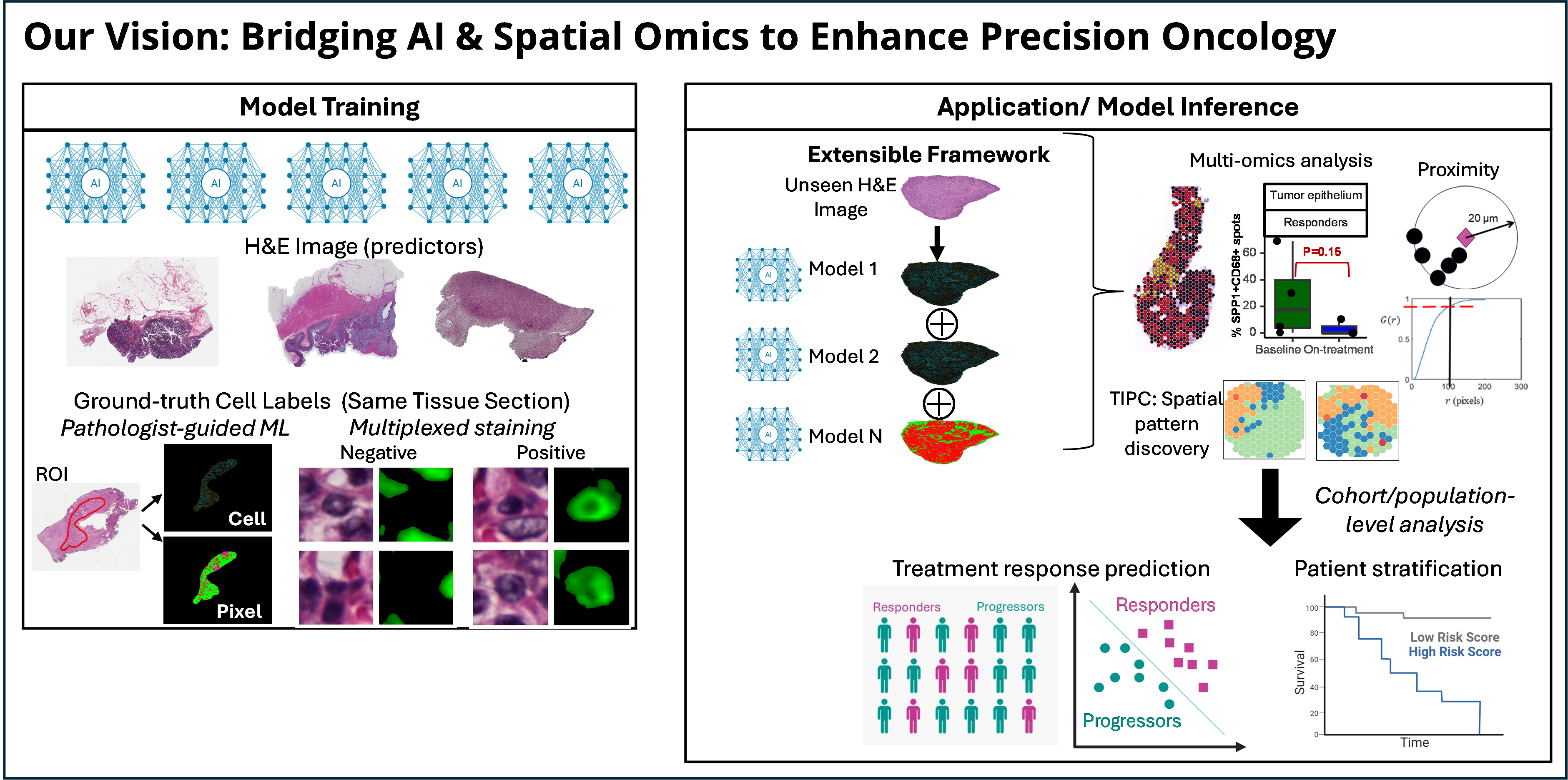
To address these challenges, we introduce a H&E-based spatial virtual Multi-Omics (svMO) Approach, utilizing virtual staining to enable large-scale, population-level tumor microenvironment studies and multi-omics biomarker development (Figure 1). Motivated by the long-standing success of H&E-based predictions, such as microsatellite instability status prediction in gastrointestinal cancer (10.1038/s41591-019-0462-y) and platinum response prediction in serous ovarian cancer (10.1186/s12916-020-01684-w), our team is advancing cellular molecular predictions to unravel complex cell-to-cell interactions and immune-to-tumor interactions crucial for tumor progression or treatment response.

We have significantly contributed to this field by ensuring precise cell label ground truth, as demonstrated in our recent work comparing cell labels generated from the same versus serial tissue sections in predicting CD3+ T-cells in lung cancer cohorts (10.3389/fimmu.2024.1404640). We also demonstrated the prognostic value of our virtual staining model tested on a public lung cohort (Figure 2). Additionally, we presented a comprehensive computational workflow for precise image registration to ensure accurate cell label ground truth (10.1136/jitc-2023-SITC2023.1282) (Figure 3). To enhance the clinical impact of our virtual staining models, we have developed a web-based visualization tool that integrates high-resolution H&E images with (virtual) molecular data. This tool was created in collaboration with colleagues from BII and IMCB. It can be accessed at https://mspc.bii.a-star.edu.sg/minhn/he2_COMET.html.

BII_Research_CIID-ASOQGA-Figure1

Figure 1. The H&E-based Multi-Spatial-Virtual-Omics (MSVO) Approach enables spatial multi-omics analysis and biomarker development using large-scale public H&E databases through virtual staining.

BII_Research_CIID-ASOQGA-Figure2

Figure 2. Training immunophenotyping virtual staining models using the same-section ground truth cell label derivation method enhances the accuracy of molecular signal prediction.

BII_Research_CIID-ASOQGA-Figure3

Figure 3. Precise cell label derivation achieved through optimized image registration and cell segmentation procedures.


Members

 Senior Scientist LAU Mai Chan   |    [View Bio]  
 Research officer  TAN Wei Kit 


Selected Publications


  1. Azam AB, Wee F, Väyrynen JP, Yim,WWY, Xue,YZ, Chua,BL, Lim,JCT, Somasundaram,AC, Tan,DSW, Takano,A, Chow,CY, Khor,LY, Lim,TKH, Yeong,J, Lau,MC, Cai,Y. Training immunophenotyping deep learning models with the same-section ground truth cell label derivation method improves virtual staining accuracy. Frontiers in Immunology, 2024. 15.

  2. Lau MC, Yi Y, Goh D, Cheung CCL, Tan B, Lim CTJ, Joseph CR, Wee F, Lee NJ, Lim X, Lim CJ, Leow WQ, Lee JY, Ng CYC, Bashiri H, Cheow PC, Chan CY, Koh YX, Tan TT, Kalmimuddin S, Tai WMD, Ng JL, Low GHJ, Lim KHT, Jin L, Yeong PSJ. Case Report: Understanding the Impact of Persistent Tissue-localization of SARS-CoV-2 on Immune Response Activity via Spatial Transcriptomic Analysis of Two Cancer Patients with COVID-19 Co-Morbidity. Front Immunol. 2022 Sep 12;13:978760. doi: 10.3389/fimmu.2022.978760. PMID: 36172383; PMCID: PMC9510984.

  3. Zhang Y, Lee RY, Tan CW, Guo X, Yim WWY, Lim JCT, Wee FYT, Wu Y, Kharbanda M, Lee JYJ, Ngo NT, Leow WQ, Loo LH, Lim TKH, Sobota RM, Lau MC, Davis MJ, Yeong J. Spatial omics techniques and data analysis for cancer immunotherapy applications. Curr Opin Biotechnol. 2024: 103111. ISSN 0958-1669. doi:10.1016/j.copbio.2024.103111.

  4. Lee RY, Ng CW, Rajapakse MP, Ang N, Yeong J, #Lau MC. The promise and challenge of spatial omics in dissecting tumour microenvironment and the role of AI. Front Oncol, 13, 1837. (IF 6.244; 0 citations)

  5. Lee RY, Wu Y, Goh D, Tan V, Ng CW, Lim JC, #Lau MC, Sheng JY. Application of Artificial Intelligence to In Vitro Tumor Modeling and Characterization of the Tumor Microenvironment. Adv. Healthc. Mater. 2023 Apr 15:2202457. (IF 11.092; 0 citations)

  6. Väyrynen JP, Haruki K, Lau MC, Väyrynen SA, Ugai T, Akimoto N, Zhong R, Zhao M, Dias Costa A, Borowsky J, Bell P, Takashima Y, Fujiyoshi K, Arima K, Kishikawa J, Shi S, Twombly TS, Song M, Wu K, Chan AT, Zhang X, Fuchs CS, Meyerhardt JA, Giannakis M, Ogino S, Nowak JA. Spatial organization and prognostic significance of NK and NKT-like cells via multimarker analysis of the colorectal cancer microenvironment. Cancer Immunol. Res. 2022;10(2):215-227.

  7. Borowsky J, Haruki K, *Lau MC, Dias Costa A, Väyrynen JP, Ugai T, Arima K, da Silva A, Felt KD, Zhao M, Twombly TS, Fujiyoshi K, Väyrynen SA, Hamada T, Mima K, Bullman S, Ng K, Meyerhardt JA, Song M, Giovannucci EL, Wu K, Zhang X, Freeman GJ, Huttenhower C, Garrett WS, Chan AT, Leggett BA, Whitehall VLJ, Walker N, Brown I, Bettington M, Nishihara R, Fuchs CS, Lennerz JK, Giannakis M, Nowak JA, and Ogino S. Association of Fusobacterium nucleatum with Specific T Cell Subsets in the Colorectal Carcinoma Microenvironment. Clin Cancer Res. 2021;27(10):2816-2826. (IF 13.801; 33 citations)

  8. Väyrynen JP, Haruki K, Väyrynen SA, *Lau MC, Dias Costa A, Borowsky J, Zhao M, Ugai T, Kishikawa J, Akimoto N, Zhong R, Shi S, Chang T-W, Fujiyoshi K, Arima K, Twombly TS, Da Silva A, Song M, Wu K, Zhang X, Chan AT, Nishihara R, Fuchs CS, Meyerhardt JA, Giannakis M, Ogino S, Nowak JA. Prognostic significance of myeloid immune cells and their spatial distribution in the colorectal cancer microenvironment. J Immunother Cancer. 2021;9(4): e002297. (IF 12.485; 12 citations)

  9. Väyrynen JP, Haruki K, *Lau MC, Väyrynen SA, Zhong R, Dias Costa A, Borowsky J, Zhao M, Fujiyoshi K, Arima K, Twombly TS, Kishikawa J, Gu S, Aminmozaffari S, Shi S, Baba Y, Akimoto N, Ugai T, Da Silva A, Guerriero JL, Song M, Wu K, Chan AT, Nishihara R, Fuchs CS, Meyerhardt JA, Giannakis M, Ogino S, and Nowak JA. The prognostic role of macrophage polarization in the colorectal cancer microenvironment. Cancer Immunol Res. 2021;9(1):8-19. (IF 12.020; 65 citations)

  10. Arima K, Lau MC, Zhao M, Haruki K, Kosumi K, Mima K, Gu M, Väyrynen JP, Twombly TS, Baba Y, Fujiyoshi K, Kishikawa J, Guo C, Baba H, Richards WG, Chan AT, Nishihara R, Meyerhardt JA, Nowak JA, Giannakis M, Fuchs CS, Ogino S. Utility of Metabolic Profiling of Formalin-Fixed Paraffin-Embedded Tissue Materials in Colorectal Cancer. Mol Cancer Res. 2020;18(6):883-890.

  11. Väyrynen JP, *Lau MC, Haruki K, Väyrynen SA, Dias Costa A, Borowsky J, Zhao M, Fujiyoshi K, Arima K, Twombly TS, Kishikawa J, Gu S, Aminmozaffari S, Shi S, Baba Y, Akimoto N, Ugai T, da Silva A, Song M, Wu K, Chan AT, Nishihara R, Fuchs CS, Meyerhardt JA, Giannakis M, Ogino S, Nowak JA. Prognostic significance of immune cell populations identified by machine learning in colorectal cancer using routine hematoxylin & eosin stained sections. Clin Cancer Res. 2020; 26(16):4326-4338. (IF 13.801; 30 citations)

  12. Melissa Zhao, *Lau MC, Haruki K, Väyrynen J, Gurjao C, Väyrynen S, Dias Costa A, Borowsky J, Fujiyoshi K, Arima K, Hamada T, Lennerz J, Fuchs C, Nishihara R, Chan A, Ng K, Zhang X, Meyerhardt J, Song M, Wang M, Giannakis M, Nowak J, Yu KH, and Ugai T, Ogino S. Bayesian risk prediction model for colorectal cancer mortality through integration of clinicopathologic and genomic data. NPJ Precis. Oncol. 7(1), 57.